Mosaicism (genetics)
Mosaicism: the coexistence of two or more genetically distinct cell lines in one individual from a single fertilized egg — causes, types, history, detection, and clinical importance.
Overview
Mosaicism, in genetics, describes a condition in which an individual contains two or more genetically distinct populations of cells that all originated from a single fertilized egg. In practice this means different genotypes exist side by side within the same body. The origin of those differences is postzygotic: they arise after formation of the zygote rather than from fusion of two separate embryos.
Image gallery
3 Images

Causes and mechanisms
Mosaicism can result from several molecular events, most of which happen during cell division or early development. Common mechanisms include:
- Mitotic recombination or crossing-over occurring during mitosis, which can shuffle alleles between sister chromatids.
- Postzygotic mutation in a single gene, producing a clone of cells with a new sequence variant.
- Chromosomal changes such as gains, losses, or structural rearrangements that affect only a subset of cells.
- Epigenetic processes such as random X chromosome inactivation in female mammals, which creates functional mosaicism between cells where different Xs are active.
Types and examples
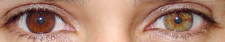
Two broad categories are often distinguished: somatic mosaicism, where the variation affects body tissues and can influence phenotype, and germline (or gonadal) mosaicism, where reproductive cells carry changes that can be passed to offspring even if the parent shows no signs. Mosaic patterns in skin or pigmentation often follow developmental cell migration and can appear as streaks or patches reflecting clonal expansion. Some inherited disorders show variable severity when mosaicism limits the fraction of affected cells.
History and discovery
The concept of recombination outside the germline was demonstrated in the 1930s. Geneticist Curt Stern showed that recombination—commonly associated with meiosis—can also take place during mitosis, producing somatic mosaics. This work established that genetically distinct cell lineages could arise within a single organism during development.
Clinical significance and detection
Mosaicism has practical implications for diagnosis, prognosis, and genetic counseling. Depending on which tissues contain the altered cells and the proportion affected, a mutation may cause disease, modify its severity, or remain clinically silent. Detecting mosaicism can require targeted approaches: cytogenetic analysis for large chromosomal abnormalities, sensitive sequencing methods to identify low-level variants, or sampling of multiple tissues because findings in blood do not always reflect other cells. Counseling must account for the possibility of germline mosaicism when estimating recurrence risk for offspring.
Distinctions and notable facts
Mosaicism is distinct from chimerism, which arises when two separate zygotes contribute genetically distinct cell lines to the same individual. Because mosaicism originates from a single fertilized egg, its pattern reflects the timing and lineage of the mutational event: earlier events produce broader involvement, later events produce more restricted patches. While sometimes neutral, mosaicism is an important source of biological diversity and a recognized mechanism in developmental conditions, cancer evolution, and variable expression of inherited diseases.
For further reading and resources, see linked topic pages on genotype, mitosis, and related subjects such as chromosomal abnormalities and X inactivation (chromosomal, X chromosome).
Questions and answers
Q: What is a mosaic in genetics?
A: A mosaic in genetics refers to the presence of two different genotypes in an individual which developed from a single fertilized egg.
Q: How does an individual develop multiple genotypes?
A: An individual develops multiple genotypes due to mosaicism, which may result from crossing-over during mitosis, a gene mutation or a chromosomal mutation during development, or X-inactivation in female mammals.
Q: What is X-inactivation in female mammals?
A: X-inactivation is a phenomenon where one of the two X chromosomes in female mammals is randomly switched off in cells, resulting in the expression of only one of the X chromosomes.
Q: Who discovered the phenomenon of mosaicism?
A: The phenomenon of mosaicism was discovered by Curt Stern, who demonstrated in 1936 that recombination, which is normal in meiosis, can also take place in mitosis, resulting in somatic mosaics.
Q: What are somatic mosaics?
A: Somatic mosaics are organisms that contain two or more genetically distinct types of tissue.
Q: What are the causes of mosaicism?
A: Mosaicism can be caused by crossing-over during mitosis, a gene mutation or a chromosomal mutation during development, or X-inactivation in female mammals.
Q: What is the significance of mosaicism in genetics?
A: Mosaicism is significant in genetics because it can result in individuals with a wide range of phenotypes, which can have important implications in fields such as medicine and evolutionary biology.
Related articles
Author
AlegsaOnline.com Mosaicism (genetics) Leandro Alegsa
URL: https://en.alegsaonline.com/art/66818
Sources
- doi.org : 10.1126/science.1092727
- pubmed.ncbi.nlm.nih.gov : 14671313
- content.nejm.org : "X Inactivation in females with X-linked disease"
- doi.org : 10.1056/NEJM199801293380511
- pubmed.ncbi.nlm.nih.gov : 9445416
- ncbi.nlm.nih.gov : ncbi.nlm.nih.gov/pubmed/21496937
- ncbi.nlm.nih.gov : "Detectable clonal mosaicism and its relationship to aging and cancer"
- doi.org : 10.1038/ng.2270
- pubmed.ncbi.nlm.nih.gov : 22561519