Mimivirus
Mimivirus is an unusually large virus first isolated from amoebae. It challenged definitions of viruses with its size, complex genome, and cellular-like features and influenced studies of viral evolution.
Overview. Mimivirus describes a group of very large viruses first identified in the early 1990s and formally described in the 2000s. It was originally discovered inside the amoeba Acanthamoeba polyphaga, where the particle was so large that it was initially misidentified as a bacterium after Gram staining. That microscopy error reflected one of the virus's notable traits: a physical size and staining behavior that can resemble small cellular microbes rather than typical viruses. Scientists often use virus to describe its biological category while emphasizing its exceptional features.
Image gallery
5 Images


Physical characteristics and genome
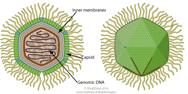
Mimivirus particles have a sizeable protein shell (capsid) decorated with a layer of fibers. The capsid itself has been an object of study because of its large diameter and unusual architecture compared with most viruses. Technical descriptions refer to the viral capsid and note measurements such as diameter when comparing it to other large nucleocytoplasmic DNA viruses. Inside the virion lies a complex double-stranded DNA genome that encodes a broad repertoire of genes, including many never before seen in viruses. Genomic surveys highlighted a richness of genetic material and prompted detailed analyses under the topic of the viral genome.
Taxonomy and related groups
Taxonomically, the organism has been treated in different ways. Some researchers describe a single species, Acanthamoeba polyphaga mimivirus, while others use the name Mimiviridae or related informal groupings to indicate a family or collection of related, giant viruses. The term genus is used in formal classification schemes, but the rapid discovery of additional giant viruses has required ongoing taxonomic revision as relationships are reassessed by genome comparisons and phylogenetic methods.
History and scientific significance
The first isolate drew attention because it blurred the line between viruses and cellular life: large size, complex gene content, and the presence of genes involved in translation and metabolism normally associated with cellular organisms. For a time it was the largest known virus until still larger examples were reported. Its discovery stimulated debate about viral origins, the limits of viral complexity, and methods used to detect microbes in environmental and clinical samples.
Ecology, research uses, and implications
Mimivirus and related giant viruses are mainly found in amoebae and other protists in aquatic or soil environments, where they can influence microbial community dynamics. Researchers study these viruses to understand virus–host interactions, the evolution of large DNA viruses, and the interplay between viruses and the microbial loop. Laboratory work uses amoebal culture systems to isolate and characterize new strains, and genomic sequencing to trace gene content and evolutionary history.
Notable facts and distinctions
- Its name derives from “mimicking microbe,” a reference to its microbe-like appearance under light microscopy.
- The initial Gram-stain confusion emphasized how morphology alone can mislead microbial identification.
- Mimivirus prompted renewed attention to giant viruses as a biologically and evolutionarily important group, distinct from classical small viruses.
- For further technical resources and comparative studies see literature linked to terms such as virus, genome, and structural topics like the capsid or measured diameter.
Readers seeking introductory reviews or primary descriptions can consult overviews that discuss isolation from Acanthamoeba, staining and identification issues tied to Gram-positive reagents, and taxonomic treatments that use terms such as genus and specific species names like Acanthamoeba polyphaga mimivirus for detailed study.
Questions and answers
Q: What is Mimivirus?
A: Mimivirus is an extra-large type of virus with a complex and large genome.
Q: How was APMV discovered?
A: APMV was discovered accidentally inside the amoeba Acanthamoeba polyphaga and seen in a gram stain, where it was thought to be a gram-positive bacterium.
Q: What is the full name of APMV?
A: APMV's full name is Acanthamoeba polyphaga mimivirus.
Q: What is Mimivirus commonly referred to as?
A: Mimivirus is commonly referred to as "mimivirus" in speech.
Q: Was Mimivirus the largest virus known until recently?
A: Yes, until October 2011 when Megavirus chilensis was discovered, Mimivirus was the largest known virus in terms of capsid diameter.
Q: How did Mimivirus get its name?
A: Mimivirus was named "mimicking microbe" due to its large size and Gram-staining properties.
Q: Is Mimivirus a single species or a group of related viruses?
A: Mimivirus is either a single species, Acanthamoeba polyphaga mimivirus (APMV), or a group of phylogenetically-related large viruses (MimiN).
Related articles
Author
AlegsaOnline.com Mimivirus Leandro Alegsa
URL: https://en.alegsaonline.com/art/65123
Sources
- doi.org : 10.1016/S0140-6736(05)62701-8
- pubmed.ncbi.nlm.nih.gov : 9093261
- virologyj.com : virologyj.com/content/2/1/62
- telegraph.co.uk : "World's biggest virus found in sea off Chile"
- nature.com : "Discovery of the giant mimivirus"