Meninges
Meninges - shortened plural of meninx encephali (from the ancient Greek μῆνιγξ méninx, German 'skin'; ἐγκέφαλος enképhalos, German 'brain', 'belonging to the brain') - are the connective tissue layers that surround the brain inside the skull (intracranially). They were first described by Herophilos of Chalcedon around 300 BC.
Three meninges are distinguished. The dura mater encephali is the hard meninges, also called pachymeninx encephali (ancient Greek παχύς pachýs, German 'coarse'). It has the arachnoid encephali (cobweb skin) more or less directly attached to it. The innermost layer is the pia mater encephali (delicate meninges). Between the arachnoid and the pia mater is the subarachnoid space (spatium subarachnoideum). These two inner meninges are also grouped together as the soft meninges, called leptomeninx encephali (Gr. λεπτός leptós, German 'light', 'fine').
Outside the skull (extracranially), the meninges continue as so-called spinal meninges, which surround the part of the central nervous system (CNS) located there. According to their anatomical affiliation with the spinal cord, they are now called dura mater spinalis, arachnoida spinalis and pia mater spinalis (Latin spinalis 'belonging to the spinal column/spinal cord').

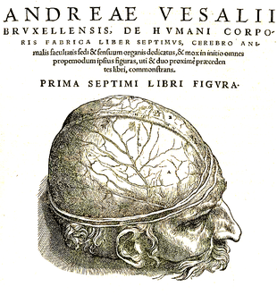
The vascular dura mater in an illustration by Andreas Vesalius (1543)
Dura mater
The dura mater (often just called "dura") is the outermost meninges. It consists of two sheets, whereby at least in the area of the skull bone the outer sheet is identical with the periosteum. The two sheets separate from each other at circumscribed points to form so-called "sinuses", a special form of venous blood conduit. The inner leaf of the dura mater draws into the great fissure between the two cerebral hemispheres and into the fissure between the two cerebellar hemispheres, and forms the falx cerebri, the falx cerebelli, and the tentorium cerebelli by juxtaposition with the inner dural leaf of each adjacent part of the brain. The falx cerebri separates the two cerebral hemispheres in the sagittal plane and merges into the falx cerebelli, which separates the two cerebellar hemispheres. Correspondingly, the tentorium cerebelli (tent roof of the cerebellum) forms, separating the cerebrum from the cerebellum. It is a structure lying rather horizontally in the skull, which has a complex three-dimensional shape due to its points of attachment to different parts of the bony skull. The diaphragma sellae forms analogously. The pituitary gland lies underneath and is thus positioned extradurally (outside the dura).
In the area of the spinal cord, the dura mater is not connected to the spinal canal. There are only a few bony attachment points of the dura mater spinalis: on the one hand, the beginning at the edge of the foramen magnum of the occipital bone, on the other hand, the end of the dural tube at the level of the 1st/2nd cruciate vertebra. The filum terminale, which begins as a continuation of the medullary cone (conus medullaris), continues from here as filum terminale externum or durale and ends at the second coccygeal vertebra, where it is also fixed bony and is in continuity with the ligamenta sacrococcygea anteriora.
In the spinal canal there is a space between the dura and the spinal canal, the epidural or peridural space, which is filled with fatty tissue. The exiting nerve roots can be anaesthetised via an injection into this space (peridural anaesthesia).
The dura is very sensitive to pain. In the cranial region, sensory innervation is provided by the ramus tentorii of the ophthalmic nerve and the rami meningei of the anterior ethmoidal nerve, the maxillary nerve, the mandibular nerve and the vagus nerve.
The dura mater is predominantly taut, collagen-fibrous connective tissue and has primarily the function of an organ capsule.

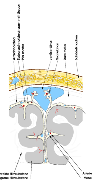
Schematic of the meninges (size ratios not shown proportionally!)
Arachnoid
The arachnoid membrane (also called cobweb skin, spider web skin or spider tissue skin) is the middle meninges of the brain. It lies adjacent to the dura mater on the inside and is separated from it by a usually closed, partly fluid-filled capillary area, the subdural fissure (spatium subdurale), which only becomes dilated and recognizable as a subdural space by excessive pathological fluid or air accumulation. This may occur when the bridging veins (venae superiores cerebri), which open into the superior sagittal sinus and thus breach the dura mater overlying the arachnoid, rupture and the escaping blood fills the subdural space. Hemorrhage into the subdural space is called a subdural hematoma (SDH).
The arachnoid, like the dura mater, skips the furrows of the brain and gets its name from the strong whitish patterning with fine fibers of collagenous connective tissue, which gives it a spider-web-like appearance. Button-shaped outpouchings protrude from the arachnoid into the venous blood conduits in the dura mater (sinus durae matris). These projections are called arachnoid villi (pacchioni granulations, granulationes arachnoideae) and are a site of CSF reabsorption.
Below the arachnoid lies the subarachnoid space (spatium subarachnoideum), which is filled with cerebrospinal fluid and thus represents the outer cerebrospinal fluid space. In this space between the arachnoid and the pia mater lie numerous superficial arteries and veins of the brain.
Questions and Answers
Q: What are the meninges?
A: The meninges are the membranes that surround and protect the brain and the spinal cord.
Q: How many layers do the meninges have in mammals?
A: The meninges have three layers in mammals: the dura mater, the arachnoid mater, and the pia mater.
Q: What is the subarachnoid space?
A: The subarachnoid space is the space between the arachnoid mater and the pia mater, where cerebrospinal fluid (CSF) is found.
Q: What is cerebrospinal fluid?
A: Cerebrospinal fluid (CSF) is a special fluid that bathes the brain and spinal cord, found in the subarachnoid space.
Q: How do the meninges and CSF work together?
A: The meninges and the CSF work together to cushion and protect the central nervous system (the brain and spinal cord).
Q: Why is CSF important?
A: CSF is important because it bathes the brain and spinal cord, providing cushioning and protection to the central nervous system.
Q: Why do the meninges have multiple layers?
A: The meninges have multiple layers to provide different levels of protection to the brain and spinal cord.
Search within the encyclopedia