Light Microscope: Principles, Components, History, and Uses
An introduction to the light microscope: how it works, main parts and optics, historical development, common types and applications, and practical limitations.
Overview
A light microscope is an optical instrument that uses visible light and lenses to enlarge small objects for observation. It operates on the same basic refracting principles as a simple telescope, but with the specimen placed very close to the objective lens so that a magnified, real image is produced and then viewed through an eyepiece. For a short comparison, see a refracting telescope discussion. Modern instruments range from basic student models to advanced laboratory systems incorporating specialized illumination and digital imaging.
Image gallery
10 Images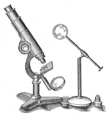


Main parts and basic operation
Typical components include an objective lens near the sample, an eyepiece or ocular lens for viewing, a stage to support the specimen, a light source and condenser to control illumination, focusing mechanisms, and sometimes a mirror to direct external light. Common items handled on the microscope are tiny organisms or cells placed on a flat glass slide. The slide is secured on the stage with clips and the stage can be moved or tilted so different portions or focal planes of the specimen can be inspected.
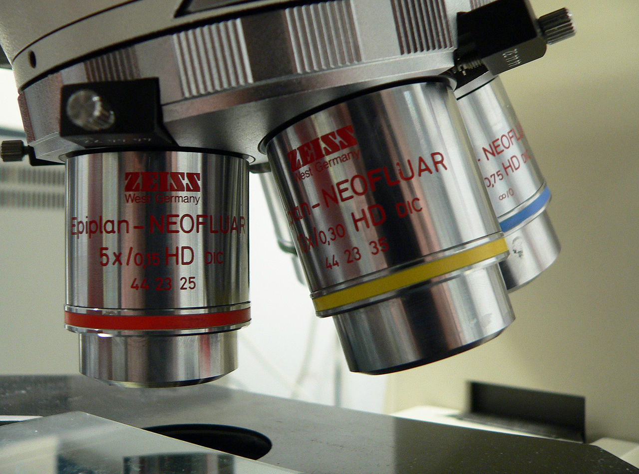
- Objective lens: the primary magnifying element located just above the specimen — see a note on objective lenses.
- Specimen and slide: samples such as a small organism are mounted on a glass slide or other support to keep them flat and accessible; many biological samples are described as microscopic organisms.
- Stage and focus: the mechanical stage holds and positions the slide; coarse and fine focus knobs bring different layers into sharp view (stage movement).
- Illumination: a built-in lamp or an adjustable mirror directs light through the condenser and specimen; historical and simpler models use a mirror to reflect light (mirror).
Optics, magnification and imaging
The overall magnification of a compound light microscope is the product of the objective and the eyepiece magnifications. Objectives produce a magnified intermediate image that the eyepiece further magnifies. Some contemporary microscopes replace or supplement the eyepiece with a sensor and live-view output, effectively turning the instrument into a digital camera for microscopy — see digital microscope systems. Typical educational microscopes include a set of objectives that allow low, medium and high magnification steps; simplified classroom models often provide basic steps that are sufficient to observe larger cells and basic tissue structures.
History and development
The development of the light microscope began in the late 16th and 17th centuries when craftsmen and natural philosophers combined small lenses to make early magnifiers. Pioneers such as Robert Hooke and Antonie van Leeuwenhoek used early microscopes to reveal previously unseen structures and organisms, laying foundational observations for cell theory and microbiology. Since that era, improvements in lens design, illumination, and sample preparation have greatly increased contrast, clarity and practical usefulness.
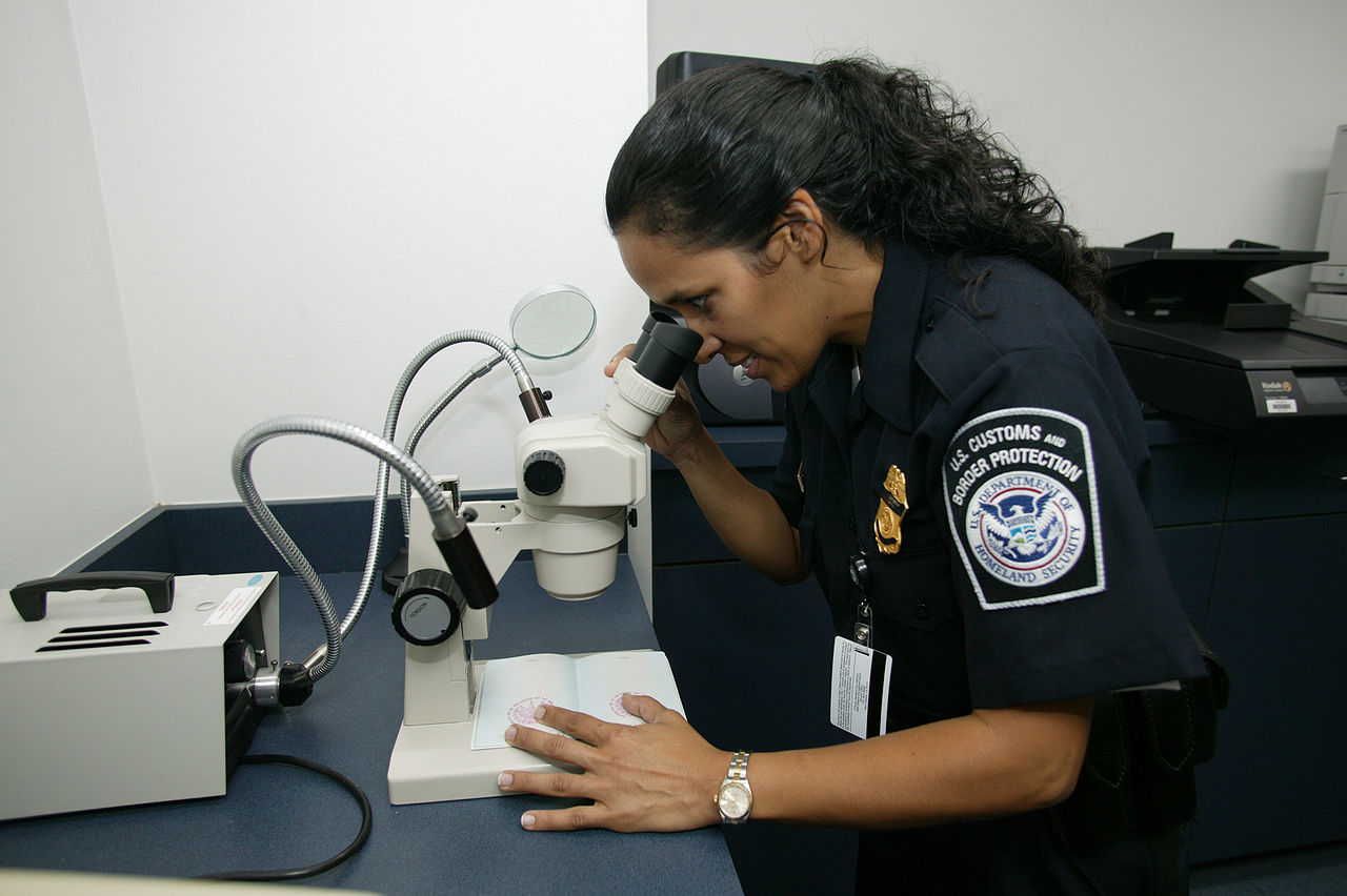
Uses, types and examples
Light microscopes are used across education, research, medicine and industry. Classroom instruments in colleges and high schools are designed for teaching and routine observation, while laboratory-grade instruments support research, clinical diagnosis and materials inspection. Examples include stained tissue slides to study cells, live-cell observation for basic biological experiments, and routine clinical smears. For typical educational contexts see college and high school resources; simple demonstrations often show basic cells and structures (cell observation).
- Compound microscopes for high magnification of thin specimens.
- Stereomicroscopes (dissecting microscopes) for low magnification and three-dimensional viewing.
- Specialized light microscopes — phase contrast, darkfield and fluorescence — which enhance contrast or reveal specific labeled structures.
Limitations and notable facts
Light microscopes are limited by the wavelength of visible light: there is a practical upper bound to the resolving power, so structures smaller than a fraction of a micrometer cannot be resolved clearly. For those scales, electron or scanning probe techniques are used. Nonetheless, light microscopy remains indispensable for visualizing cells, tissues and many microorganisms, and it continues to evolve with advances in optics, dyes, and digital imaging.
Questions and answers
Q: How does a light microscope work?
A: A light microscope works like a refracting telescope with the object being very close to the objective lens.
Q: What is a slide used for in a light microscope?
A: A slide is used to hold the object being studied, for example a tiny organism.
Q: How is the slide held in place on the microscope's stage?
A: The clips on the microscope's flat stage hold the slide in place.
Q: What does adjusting the stage of a microscope allow the user to do?
A: Adjusting the stage allows the user to add more light and to allow different layers of the object to be in focus.
Q: How does the mirror at the bottom of a microscope play a role in observing an object?
A: The mirror reflects light rays up to the object through a hole in the stage.
Q: What is the function of objective lenses in a microscope?
A: Objective lenses magnify the image of the object being observed.
Q: What magnification range is available for a light microscope commonly used in colleges and high schools?
A: The top magnification for many microscopes used in colleges and high schools is 40x, with the option of having 4x and 8x.
Related articles
Author
AlegsaOnline.com Light Microscope: Principles, Components, History, and Uses Leandro Alegsa
URL: https://en.alegsaonline.com/art/57936