
Karyotype
A karyotype is the complete set of chromosomes of an organism as seen and arranged for study; it informs chromosome number, structure, and clinical or evolutionary changes.
Overview
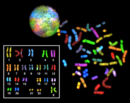
A karyotype is the complete complement of chromosomes in the nucleus of a cell, described by their number, sizes and visible features when viewed under the microscope. In biology the word applies both to the chromosome set from an individual and to the characteristic chromosome complement of a species. Analysts arrange photographed or digitally captured chromosomes into a standard display called a karyogram or idiogram to compare homologous pairs and detect abnormalities. Many foundational concepts associated with karyotypes—such as the nature of chromosomes, the cell nucleus, and the distinction between eukaryote and prokaryote cells—help explain why karyotyping is limited to organisms with visible linear chromosomes.
Image gallery
9 Images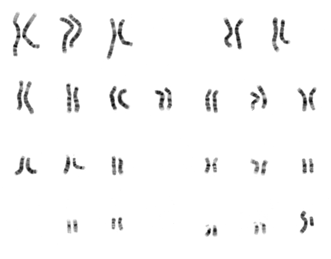



Key characteristics and terminology
Karyotypes record several features: the total chromosome count, relative lengths, position of the centromeres, and the appearance of sex chromosomes. Cytogeneticists commonly describe the somatic (body) chromosome complement as 2n and gametes or haploid cells as n. For example, most human somatic cells are diploid with 2n = 46, while human gametes are haploid with n = 23. Cells may also be polyploid (multiple complete sets) in some plants and certain animal tissues. The study of karyotypes sits within the wider field of cytogenetics, which overlaps both cytology and genetics.
How a karyotype is prepared
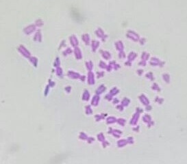
Creating a karyotype involves harvesting dividing cells, arresting them in metaphase when chromosomes are most condensed, staining, and imaging. Common staining produces banding patterns that let technicians distinguish chromosomes of similar size. A simplified workflow is:
- Collect sample from somatic tissue or cultured cells.
- Use chemical treatment to accumulate metaphase cells and apply a stain to reveal characteristic bands.
- Photograph spreads and digitally crop individual chromosomes.
- Arrange chromosome images into a karyogram by size and centromere position to pair homologues.
Clinical and biological uses
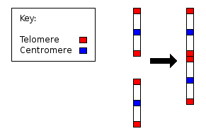
Karyotype analysis is widely used in medicine and biology. In prenatal testing it detects whole-chromosome aneuploidies (for example trisomies), large deletions or duplications, and sex-chromosome constitution by examining the complement of sex chromosomes. In oncology, changes in chromosome number and structure can indicate particular cancers or guide treatment. Comparative karyology informs evolutionary biology: differences in chromosome number and structure between populations or species can trace past events such as polyploidy and large-scale rearrangements.
History, strengths and limitations
The use of karyotypes grew in the 20th century as microscopy and staining techniques improved, establishing cytogenetics as a diagnostic and research tool. Strengths include the ability to visualize whole chromosomes and detect structural rearrangements and aneuploidies at the chromosomal scale. Limitations stem from resolution: changes smaller than the light microscope can resolve require techniques such as fluorescence in situ hybridization or genomic microarrays and sequencing. Modern practice often combines classical karyotyping with molecular methods to provide both structural and sequence-level information.
Practical distinctions and notable points
When speaking about karyotypes, it is helpful to distinguish the visual karyogram from numerical notation (for example 46,XX or 47,XY,+21 for Down syndrome). The term also intersects with taxonomy: the typical chromosome number for a given species is part of its genetic description. Specialist resources explain laboratory variations and interpretive criteria; introductory texts and databases provide standards for labeling chromosomes and describing centromere positions with reference to the resolution limits of the light microscope. For further in-depth material see basic cytogenetics primers and medical genetics references via links to educational resources and review articles (sex cell biology and gametogenesis are commonly covered together with karyotype methods).
Researchers and clinicians rely on the standardized display of chromosomes to communicate findings, whether determining an individual’s chromosomal sex, diagnosing a heritable syndrome, or documenting major rearrangements that played a role in a lineage’s evolution. As technology advances, classical karyotyping remains a foundational technique that complements molecular approaches across genetics and cell biology.
Related topics and practical guides: laboratory protocols, interpretation guidelines, and teaching materials for cytogenetics are widely available; search introductory cytogenetics literature for procedural detail and examples of karyotypes in humans and other organisms.
chromosomes • nucleus • eukaryote • species • light microscope • centromeres • sex chromosomes • cytogenetics • cytology • genetics • somatic • sex cells • haploid
Questions and answers
Q: What is a karyotype?
A: A karyotype is the number and appearance of chromosomes in the nucleus of a eukaryote cell. It describes the number of chromosomes, and what they look like under a light microscope.
Q: What is cytogenetics?
A: Cytogenetics is the preparation and study of karyotypes which combines cytology (the study of cells) and genetics (the study of heredity).
Q: What is the somatic number?
A: The somatic number refers to the basic number of chromosomes in the somatic (body) cells of an individual or species, designated 2n. For example, in humans 2n = 46.
Q: How many copies are present in normal diploid organisms?
A: In normal diploid organisms, there are two copies of each chromosome present.
Q: What does a karyogram show?
A: A karyogram or idiogram shows chromosomes arranged by size and position of centromere for chromosomes that have similar sizes. It can be used to show genetic disease, sex, diploid number, etc.
Q: How can karyotypes help identify genetic abnormalities before birth? A: Karyotypes can be studied to help identify any potential genetic abnormalities that a baby might have before it is born.
Q: How can karyotypes provide information about past evolutionary events?
A: Karyotypes may also be studied to gather information about past evolutionary events such as polyploidy (having multiple sets of chromosomes).
Related articles
Author
AlegsaOnline.com Karyotype Leandro Alegsa
URL: https://en.alegsaonline.com/art/52409