
Apoptosis: Programmed cell death in development, health, and disease
Apoptosis is the regulated process of programmed cell death essential for development and tissue balance. This article summarizes its mechanisms, roles in development and disease, detection, and how it differs from necrosis.
Overview
Apoptosis is a genetically controlled form of cell death that removes unwanted or damaged cells without provoking inflammation. Often called programmed cell death, it operates continuously throughout life to shape organs, maintain tissue quality, and eliminate cells that threaten organismal health.
Image gallery
10 Images




Key features and stages
Apoptotic cells follow a characteristic sequence of changes that distinguish them from other forms of death:
- Cell shrinkage and condensation of the nucleus (chromatin).
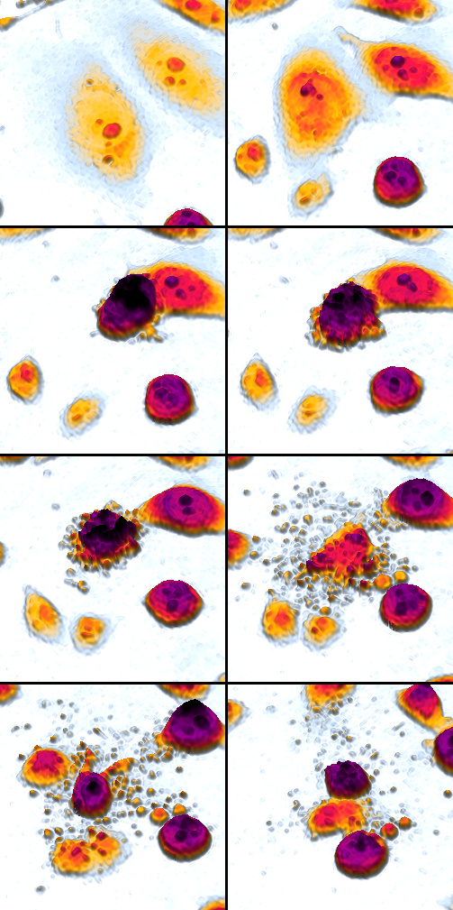
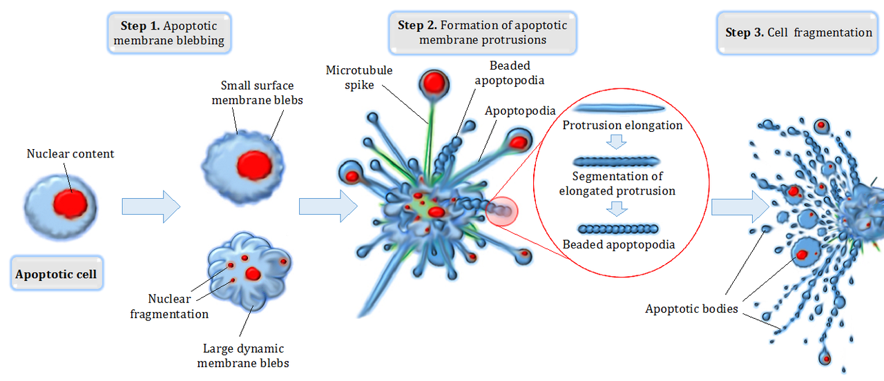
- Membrane blebbing and breakdown into membrane-bound apoptotic bodies.
- DNA fragmentation and controlled dismantling of internal structures.
- Rapid recognition and engulfment by phagocytes, preventing release of inflammatory contents.
Molecular mechanisms
Two broad pathways trigger apoptosis: an intrinsic route centered on mitochondria and an extrinsic route initiated by death receptors on the cell surface. Both converge on a family of proteases called caspases that execute the death program. Regulatory proteins such as members of the Bcl-2 family influence mitochondrial membrane integrity and determine cell susceptibility to apoptosis.
Developmental and physiological roles
Apoptosis sculpts developing tissues and removes transient structures. For example, it helps separate digits during limb formation in the embryonic limb and is active in the foetus during many stages of organogenesis. More broadly, it contributes to tissue turnover and homeostasis: rates of apoptosis are normally balanced with cell production from mitosis to maintain organ size and function. It also supports immune system development and the elimination of damaged or infected cells in adult tissues.
Clinical significance, detection, and uses
Dysregulation of apoptosis is implicated in many diseases. Insufficient apoptosis can allow cancer cells to survive, while excessive apoptosis contributes to neurodegenerative disorders and some forms of tissue wasting. Researchers and clinicians detect apoptotic cells with assays such as DNA fragmentation (TUNEL), phosphatidylserine exposure (Annexin V binding), and caspase activity tests. Therapeutic strategies aim either to promote apoptosis in cancers or to inhibit it in degenerative conditions.
Distinctions and notable facts
Apoptosis differs from necrosis: apoptosis is orderly and generally noninflammatory, whereas necrosis often results from acute injury and provokes inflammation. Historically, the concept of programmed cell death gained wide recognition in the 20th century as scientists described its role in development and disease. Its central place in biology makes apoptosis a key topic in developmental biology, immunology, and medicine. For more on tissue contexts where apoptosis is important, see tissue dynamics and on life-stage changes such as those occurring before puberty.
Further reading and resources: definition and basics, limb development, prenatal development, cell division balance, tissue remodeling, developmental timing.


Occurrence
During the development of an organism, apoptosis is essential:
- during the metamorphosis from tadpole to frog or the degeneration of the skins between the fingers/toes (interdigital skins), cells are specifically stimulated to apoptosis
- through apoptotic cell death of the cells of the vitreous body and lens of the lens eye, the light transmission of the eye lens is achieved
- to ensure the correct "interconnection" of brain structures and individual nerve cells, up to half of all originally formed nerve cells die again before birth
But it is also essential in the adult organism:
- for checking the cell count and the size of tissues
- in the rejuvenation of tissues (e.g. in the olfactory epithelium of the nose)
- in the selection and degradation of unnecessary or potentially harmful cells of the immune system
- for the elimination of degenerated cells
- to ensure plasticity in the central nervous system
- for the selection of germ cells (approx. 95 percent of germ cells are apoptotically killed before reaching maturity)
- in holocrine secretion, i.e. in the sebaceous glands of the human body
Currently, apoptosis is being researched particularly in connection with carcinogenesis and various autoimmune diseases. One goal of cancer research is to trigger controlled apoptosis in degenerated cells. However, cancer cells also use the apoptosis mechanism to switch off human defence cells, so-called tumour-infiltrating lymphocytes (TILs). Thus, an apoptosis-inducing protein, the CD95 ligand (Fas ligand), is found on the surface of various tumor cell lines. This mechanism is known as tumor counterattack.
The role of apoptosis in neurodegenerative diseases (e.g. Alzheimer's disease, Huntington's disease, Parkinson's disease, ALS) is also the subject of intense debate and a wide range of research is ongoing in this area.
Signs of apoptosis have also been found in unicellular organisms. In Saccharomyces cerevisiae (baker's yeast, brewer's yeast) - especially in old cells - various markers of apoptosis (DAPI, TUNEL staining) become visible. There is speculation about evolutionary reasons for the presence of apoptosis in unicellular organisms. One theory is that individual damaged cells sacrifice themselves and commit "suicide" for the good of the collective. This saves nutrients, which are thus available to the other cells. The goal is ultimately to preserve the genome, which is also present in virtually identical form in the other cells.
View
Histology
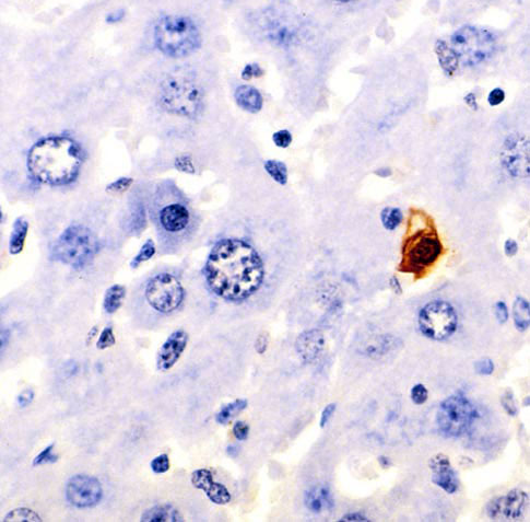
The process of apoptosis can be observed under the light microscope. First, the cell in question detaches itself from the tissue. In the further course, the cell becomes more and more eosinophilic in colour and becomes increasingly smaller. In addition, visible vesicles form on the cell membrane. The cell nucleus becomes smaller and more densely packed. In the course of apoptosis, it can also disintegrate into several parts. At the end of the process, a homogeneous eosinophilic apoptotic corpuscle remains. This is then degraded by phagocytosis. The programmed cell death does not trigger an inflammatory reaction.
Imaging techniques
Apoptosis can be detected macroscopically in vivo by imaging techniques such as positron emission tomography, fluorescence imaging and magnetic resonance imaging (molecular imaging). Modified amino acids, such as (5-dimethylamino)-1-napththalinsulfonyl-α-ethyl-fluoroalanine (NST-732) or N,N′-didansyl-L-cystine, are used as tracers.
Questions and answers
Q: What is apoptosis?
A: Apoptosis is the controlled death of a cell.
Q: What role does apoptosis play in tissue development?
A: Apoptosis plays an important role in tissue development, such as allowing digits to separate from each other during limb development in a foetus.
Q: When does apoptosis occur most often?
A: Apoptosis occurs most often until puberty begins.
Q: Is apoptosis a regulated process?
A: Yes, apoptosis is a regulated and controlled process which works throughout an organism's life cycle.
Q: How is the rate of apoptosis balanced?
A: The rate of apoptosis is balanced by the rate of cell production by mitosis.
Q: Does apoptosis happen only during certain stages of life?
A: No, apoptsis happens throughout an organism's life cycle.
Related articles
Author
AlegsaOnline.com Apoptosis: Programmed cell death in development, health, and disease Leandro Alegsa
URL: https://en.alegsaonline.com/art/4931