
Inner ear: anatomy, function, evolution and clinical importance
The inner ear is the innermost part of the vertebrate ear responsible for hearing and balance. It includes the cochlea and vestibular apparatus housed in the bony labyrinth and connects to the brain via cranial nerve VIII.
The inner ear is the deepest portion of the vertebrate auditory system and performs two essential roles: converting sound into neural signals and sensing head motion and orientation. This complex structure sits within the temporal bone and contains a system of cavities and fluid-filled channels that form a protective bony labyrinth. For general context see the vertebrate ear and how the inner ear differs among mammals.
Image gallery
9 Images
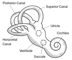

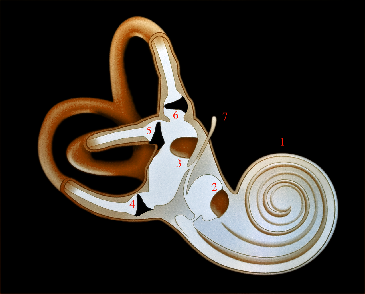

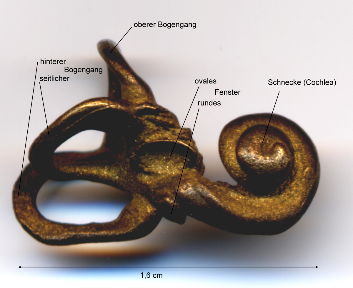

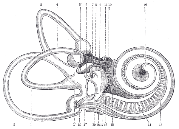
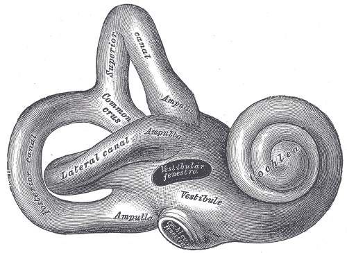
Anatomy and main parts
Anatomically the inner ear can be divided into two functional compartments: the cochlea for hearing and the vestibular apparatus for balance. The principal elements include:
- Cochlea — a spiraling, fluid-filled chamber that houses the organ of Corti, where sensory hair cells transduce pressure waves into electrical impulses. See also cochlea.
- Vestibular system — including three semicircular canals and two otolithic organs (utricle and saccule) that detect angular and linear acceleration; the canals are oriented roughly at right angles to one another to sense rotation in three dimensions (right angles).
- Bony and membranous labyrinths — the membranous labyrinth sits within the bony case and contains endolymph, while perilymph surrounds it.
How the inner ear works
Sound waves transmitted through the outer and middle ear set the cochlear fluids in motion. Movement of the basilar membrane bends hair cell stereocilia in the organ of Corti, opening ion channels and generating receptor potentials that are sent to the brain via the auditory branch of cranial nerve VIII (eighth cranial nerve). In the vestibular portion, movement of endolymph inside the semicircular canals deflects cupulae and hair cells to signal angular acceleration; otolithic membranes in the utricle and saccule respond to gravity and linear acceleration.
Evolution and comparative notes
The inner ear is present across vertebrates but shows adaptation to different lifestyles. Fish and amphibians use otoliths—small calcareous structures—to aid balance and hearing in water. Reptiles and birds have variations of the cochlear duct suited to their frequency ranges, while mammals developed a highly coiled cochlea that enhances frequency discrimination. Despite these differences, the inner ear consistently connects to the brain via the vestibulocochlear pathway.
Clinical relevance and examples
Dysfunction of the inner ear causes common problems such as sensorineural hearing loss, vertigo, tinnitus, and balance disorders. Conditions like Meniere's disease, labyrinthitis, and age-related degeneration affect either cochlear or vestibular components. Diagnostic methods include audiometry, vestibular function testing and imaging; treatments range from rehabilitation and medication to devices such as hearing aids and cochlear implants.
Notable facts and distinctions
The inner ear is encased in dense bone for protection and has a unique ionic environment (endolymph vs perilymph) that is crucial for sensory transduction. The three semicircular canals form an orthogonal set to encode rotation, while the cochlea separates sound frequencies along its length. For further anatomical detail consult summaries of the bony labyrinth, the separate description of the balance system, and sources on auditory mechanics orientation and signal transmission.
Understanding the inner ear combines anatomy, fluid mechanics, and neurophysiology; it remains a focal point for research into hearing restoration, balance rehabilitation and evolutionary biology.
Questions and answers
Q: What is the inner ear?
A: The inner ear is the innermost part of the vertebrate ear, responsible for sound detection and balance.
Q: What are the two main working parts of the inner ear in mammals?
A: The two main working parts of the inner ear in mammals are the cochlea and the balance system.
Q: What is the cochlea?
A: The cochlea is a part of the inner ear that is dedicated to hearing, converting sound pressure patterns from the outer ear into electrochemical impulses which are passed on to the brain via the auditory nerve.
Q: What is the balance system?
A: The balance system is a part of the inner ear consisting of three semi-circular canals, set at right angles to each other.
Q: In what types of animals is the inner ear found?
A: The inner ear is found in all vertebrates, with variations in form and function.
Q: What nerve is responsible for supplying the inner ear in all vertebrates?
A: The eighth cranial nerve is responsible for supplying the inner ear in all vertebrates.
Q: How does the cochlea convert sound pressure patterns into electrochemical impulses?
A: The cochlea contains tiny hair cells that are moved by the sound pressure waves. This movement triggers the release of chemicals that generate electrical signals, which are then carried by the auditory nerve to the brain.
Related articles
Author
AlegsaOnline.com Inner ear: anatomy, function, evolution and clinical importance Leandro Alegsa
URL: https://en.alegsaonline.com/art/47391