
Hyoid bone
A small U-shaped bone in the anterior neck that supports the tongue and larynx and serves as an attachment site for muscles and ligaments involved in swallowing and speech.
Overview
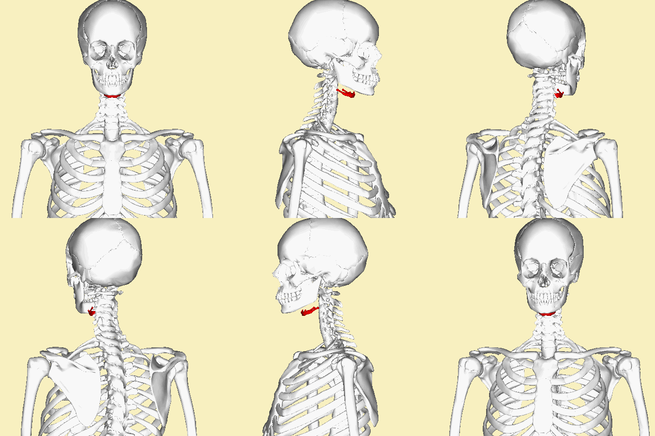
The hyoid is a small, curved bone in the front of the neck that is commonly described as horseshoe-shaped. It sits in the midline between the lower jaw and the throat, roughly between the chin (chin) and the prominence of the larynx often called the Adam's apple (Adam's apple). At rest the hyoid lies approximately at the level of the back of the lower jaw (the mandible) and near the third cervical vertebra. Unique among bones of the skeleton, the hyoid does not form a joint with any other bone and is instead suspended by soft tissues.
Image gallery
9 Images



Anatomy and relationships
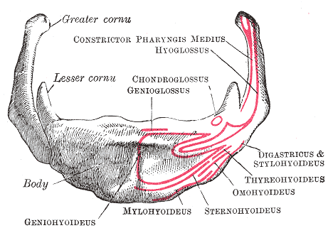
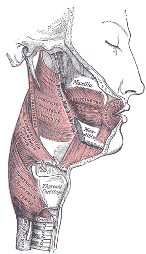
Structurally, the hyoid consists of a central body and two pairs of projections known as the greater and lesser cornua (horns). It is linked to the skull, jaw and larynx by a network of muscles and ligaments (muscles and ligaments) rather than by direct bony connections. These attachments place the hyoid at the junction of oral and pharyngeal anatomy: it relates superiorly to the tongue, inferiorly to the larynx, and posteriorly toward the pharynx and epiglottis.
Function
The hyoid acts as a mobile anchor for muscles of the tongue and floor of mouth on its superior surface and for muscles that elevate or lower the larynx on its inferior surface. This makes it central to swallowing, maintaining an open airway, and contributing to articulation during speech. Movement of the hyoid helps coordinate the sequence of events that propel food from the oral cavity into the esophagus and also adjusts the position of the larynx during breathing and phonation.
Clinical and forensic significance
Because the hyoid is suspended by soft tissues, trauma or fracture of the bone is uncommon but clinically important. Hyoid fractures can result from direct blows, compressive neck injuries, or strangulation and may be examined in forensic investigations. The bone is also a surgical landmark in procedures involving the airway, neck tumors, and reconstructive surgery. Developmentally, the hyoid originates from the pharyngeal arches and may show variation in shape, size, and degree of ossification across individuals and with age.
Key points
- Small, U-shaped bone in the anterior neck (horseshoe-shaped).
- Suspended by muscles and ligaments, not articulating with other bones.
- Serves as an attachment for the tongue, floor of mouth, and laryngeal elevators (larynx).
- Relevant in swallowing, speech, airway mechanics, surgery, and forensic examinations.
Questions and answers
Q: What is the hyoid bone?
A: The hyoid bone is a horseshoe-shaped bone located in the front of the neck, between the chin and the thyroid cartilage (Adam's apple).
Q: What is the position of the hyoid bone in the neck?
A: At rest, the hyoid bone lies at the level of the mandible in the front and the third cervical vertebra behind.
Q: Is the hyoid bone connected to other bones directly?
A: No, the hyoid bone is not directly connected to other bones.
Q: How is the hyoid bone connected to other structures?
A: The hyoid bone is connected only to muscles and ligaments.
Q: What is the function of the hyoid bone?
A: The hyoid bone aids in tongue movement and swallowing. It also provides attachment to the muscles of the floor of the mouth and the tongue above, the larynx below, and the epiglottis and pharynx behind.
Q: What are the muscles attached to the hyoid bone?
A: The hyoid bone provides attachment to the muscles of the floor of the mouth and the tongue above, the larynx below, and the epiglottis and pharynx behind.
Q: How does the hyoid bone aid in swallowing?
A: The hyoid bone moves upward and forward during swallowing, which helps to lift the larynx and close off the opening to the trachea, preventing food or liquid from entering the airways.
Related articles
Author
AlegsaOnline.com Hyoid bone Leandro Alegsa
URL: https://en.alegsaonline.com/art/46147