Heterochromia: Causes, Types, and Clinical Significance
Heterochromia is a difference in iris coloration between or within the eyes. This article explains types, causes (genetic and acquired), diagnostic considerations, occurrences in animals, and related terms.
Heterochromia describes a visible difference in eye coloration, most often involving the irises. It can be present at birth or develop later in life and ranges from complete differences between the two eyes to smaller, sectoral changes within a single iris. For general background see further reading.
Image gallery
7 Images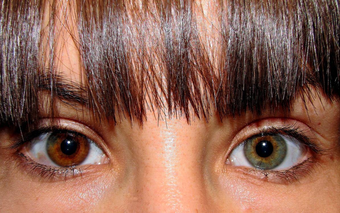


Common forms and appearance
Clinically, heterochromia is often categorized as complete, sectoral (partial), or central. Complete heterochromia means each eye has a distinct, uniform color. Sectoral heterochromia features a wedge or portion of a different color in one iris. Central heterochromia involves a ring or radial variation around the pupil. These visible patterns reflect how pigment is distributed in the iris stroma and epithelium; detailed anatomy of the iris is discussed in specialist sources such as anatomy references.
Causes and underlying mechanisms
Causes fall into two broad groups: congenital (genetic or developmental) and acquired. Congenital causes include genetic mosaicism and inherited syndromes that affect pigment cells. Mosaicism can produce localized differences in pigment because of genetic changes in cell lineages during embryonic development; see resources on embryology at embryonic development. Specific genetic conditions associated with heterochromia include certain forms of Waardenburg syndrome and other pigmentary disorders; genetics overview is available at genetics summaries.
Acquired causes and clinical significance
Heterochromia that appears later may result from eye injury, inflammation, tumors, or neurologic conditions such as Horner's syndrome. Some ocular diseases (for example, Fuchs heterochromic iridocyclitis) and prolonged use of certain glaucoma medications can alter iris pigmentation. When heterochromia is new or accompanied by other symptoms (vision change, pain, redness), medical evaluation is recommended to exclude serious causes.
Diagnosis, management, and distinctions
Diagnosis typically involves clinical examination, photographic documentation, and sometimes imaging or laboratory tests to investigate suspected underlying disease. Most benign congenital cases require no treatment; management is directed at any associated condition. In cosmetic contexts, colored contact lenses may be used, but these require appropriate fitting and care. Terminology varies: traditional Latin terms include heterochromia iridis and heterochromia iridum, while heterochromia iridium is a common grammatical error noted in some older texts (language notes).
Occurrence in animals and cultural notes
Heterochromia is also seen in several animal species, notably dogs, cats, and horses; certain breeds carry genetic variants that make it more common. It has attracted human interest for cosmetic and cultural reasons, sometimes being featured in art and media. For veterinary perspectives and breed associations, see animal resources and veterinary references.
- Key points: heterochromia is a descriptive finding, not a diagnosis.
- If new or symptomatic, seek ophthalmic assessment.
- Many cases are harmless, but some indicate underlying disease.
Questions and answers
Q: What is heterochromia?
A: Heterochromia is a condition where people or animals have different coloured eyes.
Q: What is genetic mosaicism?
A: Genetic mosaicism is a genetic cause of heterochromia that occurs when changes happen in the dividing cells leading up to iris formation in the embryo.
Q: Are there environmental causes of heterochromia?
A: Yes, environmental damage can occur to the eyes and result in heterochromia.
Q: Can various diseases and medical conditions result in changes in eye colour?
A: Yes, various diseases and medical conditions can result in changes in eye colour and cause heterochromia.
Q: Where is the condition of heterochromia most easily seen?
A: The condition of heterochromia is most easily seen in the iris of the eye.
Q: What are the different terms used for heterochromia when specifically referring to eye colour?
A: The terms Hetrochromia iridis, Heterochromia iridum (plural form), and Heterochromia iridium (a mistake in Latin grammar) are variously used when specifically referring to the eye colour in heterochromia.
Q: Can heterochromia occur in tissues other than the iris of the eye?
A: Yes, similar events may happen in other tissues, which may result in visible tissue differences.
Related articles
Author
AlegsaOnline.com Heterochromia: Causes, Types, and Clinical Significance Leandro Alegsa
URL: https://en.alegsaonline.com/art/43951