
Gram staining (Gram's method)
A microbiological staining technique that classifies bacteria as gram-positive or gram-negative based on cell wall properties; widely used for identification and guiding clinical decisions.
Overview
Gram staining, also called Gram's method, is a fundamental laboratory technique used to differentiate bacteria into two broad categories: gram-positive and gram-negative. The classification reflects differences in cell envelope structure that influence how bacterial cells interact with dyes and solvents. In clinical and research laboratories the Gram stain is usually the first test performed on a bacterial specimen because it provides rapid, useful information about organism type and likely antibiotic susceptibility.
Image gallery
6 Images



Procedure and underlying mechanism
The staining procedure normally follows a four-step sequence. First, a primary violet dye (crystal violet) stains all cells. Second, an iodine solution acts as a mordant to form a crystal violet–iodine complex. Third, a decolorizing agent (usually ethanol or acetone–alcohol) is applied; this is the critical step that differentially removes the dye complex from some cells. Finally, a counterstain such as safranin or fuchsin is added to color cells that lost the primary stain.
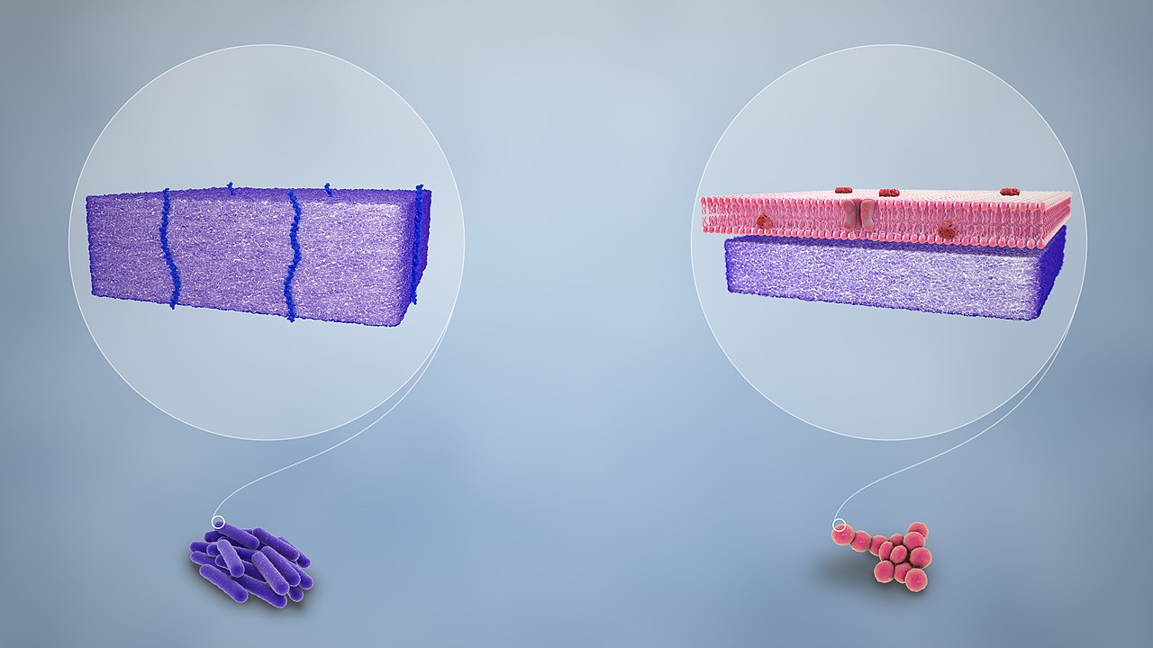
The difference in outcome is caused by structural features of the cell envelope. Gram-positive bacteria have a relatively thick peptidoglycan layer that traps the dye–iodine complex when the decolorizer dehydrates and tightens the wall. Gram-negative bacteria have a thinner peptidoglycan layer and an outer membrane containing lipopolysaccharides; the decolorizer disrupts the outer membrane and allows the complex to wash out, so these cells take up the counterstain and appear pink or red.
History and development
The technique was developed by the Danish physician Hans Christian Gram in the 1880s as a way to make bacteria in tissue samples more visible under the microscope. He described his method in 1884 while working in a hospital laboratory. Later researchers refined reagent choices and timings; the essential principles of differential staining that Gram introduced remain central to bacterial microscopy today.
Uses, examples and limitations
Gram staining guides initial diagnosis and management in medicine: for example, gram-positive cocci in clusters suggest Staphylococcus, while gram-negative rods point toward organisms such as Escherichia coli or Pseudomonas. It also informs laboratory decisions about culture media and further biochemical or molecular testing.
- Typical gram-positive groups: Staphylococcus, Streptococcus, Bacillus.
- Typical gram-negative groups: Escherichia, Pseudomonas, Neisseria.
Despite its value, the Gram stain has limits. Some bacteria are gram-variable or do not stain reliably (for example, Mycobacterium with waxy cell walls, or wall-less bacteria such as Mycoplasma). Improper technique, overly old cultures, or intracellular localization can also yield misleading results, so Gram findings are interpreted alongside clinical context and further laboratory tests.
Further reading and resources
For practical protocols, interpretations, and historical background, see these resources:
Questions and answers
Q: What is Gram staining?
A: Gram staining is a method of classifying bacteria into two large groups: gram-positive and gram-negative.
Q: Who invented Gram staining?
A: Gram staining was invented by Hans Christian Gram.
Q: How does Gram staining work?
A: Gram staining stains bacteria according to the chemical and physical properties of their cell walls. A violet dye is put on the bacteria, which stains peptidoglycan, a thick layer found only in gram-positive bacteria. After the first stain, another stain (usually safranin or fuchsin) gives all gram-negative bacteria a red or pink color.
Q: Why is the Gram stain important?
A: The Gram stain is important because it is almost always the first step in the identification of a bacterial organism.
Q: Can all bacteria be classified by Gram staining?
A: No, not all bacteria can be classified by Gram staining. Bacteria for which the method does not work are called 'gram-variable' or 'gram-indeterminate'.
Q: What did Gram use the Gram stain for initially?
A: Gram used the Gram stain to make bacteria in the lungs easier to see.
Q: When was the Gram stain method published?
A: The Gram stain method was published by Gram in 1884.
Tags
Related articles
Author
AlegsaOnline.com Gram staining (Gram's method) Leandro Alegsa
URL: https://en.alegsaonline.com/art/40113
Sources
- ncbi.nlm.nih.gov : "The Gram stain and the etiology of lobar pneumonia, an historical note"
- pubmed.ncbi.nlm.nih.gov : 13685217
- books.google.com : Milestones in Microbiology 1546–1940
- hoslink.com : "Pioneers in medical laboratory science: Christian Gram 1884"