
Golgi complex (Golgi apparatus): structure, function, and significance
Overview of the Golgi complex: its structure, roles in protein and lipid processing, historical discovery, variations across organisms, and importance in cellular transport and health.
The Golgi complex, also called the Golgi apparatus or simply the Golgi, is a membrane-bound organelle found in nearly all eukaryotic cells. It occupies a central place in the cell's endomembrane system, receiving newly made proteins and lipids from the endoplasmic reticulum and preparing them for delivery to their final destinations. The Golgi's ability to modify, sort and package macromolecules underlies secretion, membrane maintenance, and the biogenesis of lysosomes and similar organelles in diverse organisms.
Image gallery
5 Images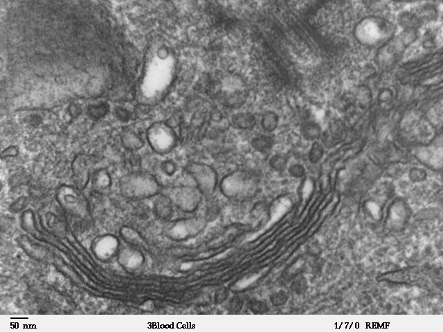



Structure and organization
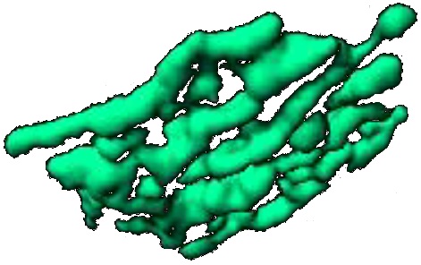
Microscopically, the Golgi is composed of flattened, stacked membrane sacs called cisternae. These stacks present a polarity: a receiving or cis face, a medial region where many chemical modifications occur, and a trans face that dispatches processed cargo. The trans side often connects to a wider area known as the trans‑Golgi network that sorts material into transport carriers. Smaller transport vesicles shuttle cargo between the Golgi and other compartments; coat proteins such as COP and clathrin help form and target these carriers. Proteins that help maintain Golgi architecture include matrix components often referred to as golgins and tethering factors.
Principal functions
The Golgi performs several interrelated tasks in the secretory pathway. Key activities include:
- Post‑translational modification: adding or trimming sugar chains (glycosylation), adding sulfate groups, or other chemical changes that affect folding and function of proteins and lipids.
- Sorting and packaging: segregating molecules destined for secretion, the plasma membrane, or endosomes/lysosomes. Many lysosomal enzymes are tagged in the Golgi for proper delivery.
- Generation of transport carriers: budding vesicles and tubules that ferry cargo to and from the Golgi and other compartments.
These operations are enzymatic and sequential, so cargo moves through distinct Golgi compartments as it matures.
Discovery and historical context
The organelle was first visualized by Italian physician-scientist Camillo Golgi in 1898 using a silver staining technique. Early skepticism dismissed his images as artifacts, but later advances—especially in electron microscopy—confirmed the Golgi's presence and revealed its detailed architecture. Over the 20th century, studies established the Golgi as a dynamic station in intracellular transport rather than a static storage compartment.
Variations, models and biological importance
Golgi structure and number vary between cell types and organisms. Animal cells often have a centralized Golgi near the nucleus, while plant cells and many protists display multiple separated stacks (sometimes called dictyosomes). Two major conceptual models describe how cargo moves through the Golgi: the cisternal maturation model and the vesicular transport model; modern evidence supports aspects of both. Disruption of Golgi function affects glycosylation and trafficking and is associated with cellular stress and a range of diseases, underscoring the organelle's physiological significance.
Examples, distinctions and further reading
Practical examples of Golgi activity include secretion of digestive enzymes, membrane protein delivery needed for cell signaling, and assembly of complex extracellular matrix components in multicellular organisms. Distinct features include the Golgi's polarized organization, its centrality to the secretory pathway, and its interactions with cytoskeletal elements that position and shape stacks. For introductions, reviews, and specialized topics see: overview articles, structural studies, cell biology texts, and resources on secretory pathways, lysosome formation, golgin proteins and membrane traffic.
Questions and answers
Q: What is the Golgi complex?
A: The Golgi complex, also known as the Golgi apparatus or simply the Golgi, is a cytoplasmic organelle found in eukaryote cells such as animals, plants, and fungi.
Q: Who discovered the Golgi complex?
A: The complex was discovered by Camillo Golgi in 1898.
Q: What does it look like?
A: It is made of several flattened sac-like membranes which look like a stack of pancakes.
Q: What is its main function?
A: The main function of the Golgi apparatus is to process and package macromolecules, such as proteins and lipids.
Q: How does it process proteins for secretion?
A: Much of the enzymatic processing is post-translational modification of proteins. The Golgi complex inspects them for flaws and discards extra material added during their manufacture, wraps them up and then targets them for packaging. It also releases special enzymes called lysosomes which clean off any extra amino acids before they are released into the cytoplasm.
Q: How does it relate to other cellular membrane systems?
A: The Golgi complex is part of the cellular membrane system, along with the endoplasmic reticulum.
Related articles
Author
AlegsaOnline.com Golgi complex (Golgi apparatus): structure, function, and significance Leandro Alegsa
URL: https://en.alegsaonline.com/art/39547
Sources
- link.springer.com : link.springer.com/article/10.1007/s00418-013-1128-3