Fallopian tube
A paired reproductive tube that carries the ovum from the ovaries to the uterus and is the usual site of fertilization; important in fertility and reproductive health.
The fallopian tubes, also called uterine tubes or oviducts, are paired ducts in the female reproductive system that link the ovaries to the uterus. They provide the route by which an oocyte leaves the ovary and travels toward the uterine cavity. In most mammals, including humans, the tubes are the typical site where an egg meets sperm and fertilization occurs.
Image gallery
9 Images


Anatomy and structure
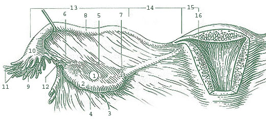
Each tube lies on one side of the uterus and opens near the corresponding ovary, while the other end joins the uterine wall at the uterus. The tubes are composed of several segments: the fimbriae and infundibulum at the ovarian end, the ampulla (a widened middle portion), the isthmus (a narrower region), and the intramural or uterine part that traverses the uterine wall. The inner lining is a mucosa with ciliated and secretory epithelial cells; surrounding smooth muscle layers generate peristaltic contractions that help propel gametes and embryos.
Function and physiology
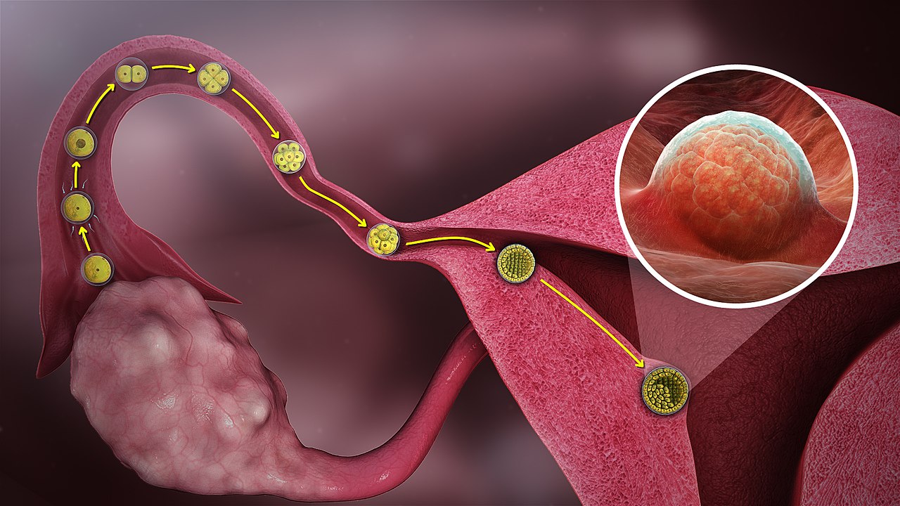
Following ovulation, fimbrial projections sweep the released ovum into the tube. Ciliary beating and coordinated muscular activity move the oocyte or early embryo toward the uterus. Sperm ascend from the uterine cavity into the tube, and most natural fertilizations occur in the ampulla when sperm meet the ovum. Tubal secretions provide a nutritive and protective environment for early embryonic development until uterine implantation.
Clinical significance
Tubal health is crucial for fertility. Damage, scarring, or blockage—commonly resulting from infections, surgery, or endometriosis—can prevent egg and sperm from meeting and is a common cause of infertility. An ectopic pregnancy, in which an embryo implants within a tube instead of the uterus, is a serious complication requiring prompt treatment. Surgical sterilization procedures (tubal ligation) intentionally interrupt tubal continuity to prevent pregnancy.
Diagnosis, treatment and prevention
- Investigation of tubal patency often uses imaging such as hysterosalpingography or laparoscopy with dye testing.
- Infections like pelvic inflammatory disease are treated with antibiotics; surgical repair is sometimes attempted for certain tubal lesions.
- Assisted reproductive technologies can bypass tubal problems when necessary.
Development and notable facts
Embryologically, the fallopian tubes develop from the paramesonephric (Müllerian) ducts. In adults they are slender ducts several centimeters long (commonly around 10–12 cm) and vary with age and reproductive history. Because they are paired and closely associated with the ovaries and uterus, their form and function are central to reproduction and are frequently discussed in fertility care, gynecologic surgery, and reproductive biology texts. For more general background see resources on the sexual and reproductive system.
Further reading and clinical guidelines are available from authoritative gynecology and reproductive medicine sources; introductory overviews of related structures can be found through general anatomy references and patient information portals (ovary information, uterus information).
Questions and answers
Q: What are Fallopian tubes?
A: Fallopian tubes are pathways that connect the ovaries to the uterus.
Q: What are some other names for Fallopian tubes?
A: Fallopian tubes are also known as oviducts and uterine tubes.
Q: What is the function of Fallopian tubes?
A: Fallopian tubes function to let the ovum pass into the uterus where they can be fertilized by sperm during sexual intercourse.
Q: How many Fallopian tubes are attached to the uterus?
A: There are two Fallopian tubes attached to either side of the uterus.
Q: Are Fallopian tubes present in males?
A: No, Fallopian tubes are not present in males.
Q: What happens to the ovum after it enters the Fallopian tubes?
A: After the ovum enters the Fallopian tubes, it can be fertilized by sperm during sexual intercourse.
Q: Can a woman become pregnant without Fallopian tubes?
A: No, a woman cannot become pregnant without Fallopian tubes as they are necessary for the ovum to be transported from the ovaries to the uterus and for fertilization to occur.
Related articles
Author
AlegsaOnline.com Fallopian tube Leandro Alegsa
URL: https://en.alegsaonline.com/art/33381
Sources
- ect.downstate.edu : 43:04-0101
- gfmer.ch : Fallopian tube
- www3.umdnj.edu : Oviduct