Ethmoid bone: structure, function, and clinical significance
Small, lightweight skull bone between the orbits that separates the nasal cavity from the cranial cavity; important for smell, the nasal septum, and the paranasal sinuses.
The ethmoid bone derives its name from the Greek ethmos, meaning "sieve," a reference to its porous appearance. It is a delicate, cubical bone situated at the roof of the nose between the two orbits, where it helps separate the nasal cavity from the cranial cavity. Located near the midline of the skull, the ethmoid contributes to the medial walls of the orbits and forms part of the roof of the nose. Its light, perforated nature is often described as spongy or sieve-like.
Image gallery
10 Images


Structure and parts
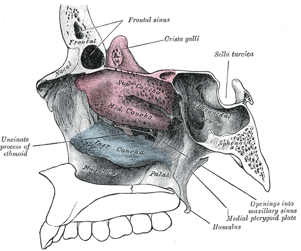
The ethmoid is commonly described in three principal regions: the cribriform plate, the perpendicular plate, and the lateral masses (ethmoidal labyrinths). Key components include:
- Cribriform plate: a horizontal, perforated portion that transmits olfactory nerve fibers.
- Crista galli: an upward bony ridge for attachment of the falx cerebri.
- Perpendicular plate: descends to contribute to the upper part of the nasal septum.
- Ethmoidal labyrinths (lateral masses): contain many air cells and form part of the medial orbital wall and lateral nasal wall, including the superior and middle nasal conchae.
Function and importance
The ethmoid plays several roles: it provides a pathway for olfactory nerves to the brain, supports the nasal septum, contributes to the structure of the paranasal sinuses via the ethmoidal air cells, and completes parts of the orbital cavities. Because the cribriform plate is thin and perforated, it is essential for the sense of smell but also vulnerable to injury that can cause cerebrospinal fluid leaks or loss of smell (anosmia).
Clinically, ethmoid fractures may accompany facial trauma and can permit communication between the sterile intracranial space and the nasal passages, risking infection. Chronic inflammation of the ethmoidal air cells (ethmoiditis) can affect breathing and sinus drainage. Surgeons performing endoscopic sinus or skull-base procedures must take care around the ethmoid to avoid damage to the olfactory fibers and the orbits.
During development the ethmoid forms from several ossification centers in the fetal skull and becomes a single midline bone. Its location and thin plates make it a central anatomical landmark in imaging and surgery of the anterior cranial base. For an anatomical overview consult sources on the skull anatomy and nasal cavity relationships; for clinical guidance see references on sinus disease and skull-base surgery (skull, cranial cavity).
Notable facts: the sieve-like cribriform plate gives the bone its name and unique vulnerability; the ethmoidal air cells are among the most variable of the paranasal sinuses; and the crista galli provides a firm anchor for the dural partition between cerebral hemispheres.
Questions and answers
Q: What is the ethmoid bone?
A: The ethmoid bone is a bone in the skull that is located at the roof of the nose and separates the nasal cavity from the brain.
Q: What does the name "ethmoid" mean and why is it called that?
A: The name "ethmoid" comes from the Greek word "ethmos," which means "sieve." The ethmoid bone is called that because it has a sieve-like structure.
Q: Where is the ethmoid bone located in the skull?
A: The ethmoid bone is located at the roof of the nose, between the two orbits.
Q: What is the structure of the ethmoid bone like?
A: The ethmoid bone has a cubical shape and is lightweight due to a spongy construction.
Q: What is the function of the ethmoid bone in the skull?
A: The main function of the ethmoid bone is to separate the nasal cavity from the brain.
Q: Why is it important for the ethmoid bone to be lightweight?
A: The ethmoid bone needs to be lightweight because it is located at the top of the nose and is required to support the skull while minimizing weight.
Q: What happens if the ethmoid bone is damaged or broken?
A: If the ethmoid bone is damaged or broken, it can lead to complications such as a loss of sense of smell or disruption of the brain.
Related articles
Author
AlegsaOnline.com Ethmoid bone: structure, function, and clinical significance Leandro Alegsa
URL: https://en.alegsaonline.com/art/32389