Endoplasmic reticulum
The endoplasmic reticulum (ER) is a membrane-bound eukaryotic organelle involved in protein and lipid synthesis, calcium storage, molecular transport, and quality control within the cell.
The endoplasmic reticulum (ER) is a continuous membrane system found in eukaryotic cells that forms flattened sacs, tubules and an internal lumen. As a central organelle, the ER provides routes for molecules to move to specific destinations rather than diffusing freely in the cytoplasm; it is often functionally and physically connected to the nuclear envelope and the secretory pathway (cytoplasm, nucleus). The ER is absent from prokaryotes and is a defining feature of eukaryotic cells compared with prokaryotic ones.
Image gallery
6 Images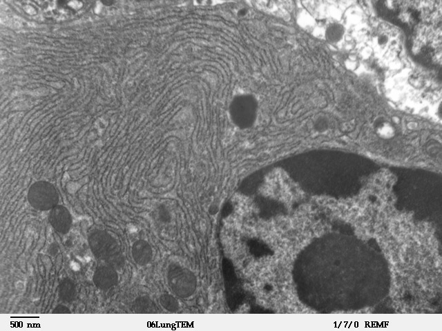


Structure and subtypes
The ER exists in two major morphologies that reflect distinct roles. The rough endoplasmic reticulum (RER) has ribosomes attached to its cytosolic surface and appears 'rough' under the electron microscope; these ribosomes translate proteins that enter the ER lumen or become membrane proteins. The smooth endoplasmic reticulum (SER) lacks bound ribosomes and forms more tubular networks involved in lipid and steroid synthesis, membrane assembly, and specialized metabolic reactions. Muscle cells contain a specialized ER called the sarcoplasmic reticulum (SR) that concentrates and releases calcium to control contraction (ribosomes, protein secretion, muscle).
Principal functions
- Protein synthesis and folding: nascent secretory and membrane proteins enter the ER, where chaperones assist folding and biochemical modifications such as glycosylation occur.
- Lipid and steroid biosynthesis: enzymes in the SER build phospholipids, cholesterol derivatives and steroid hormones that form cellular membranes and signaling molecules.
- Calcium storage and signaling: the ER lumen serves as a major intracellular calcium store; regulated release and uptake of Ca2+ control many pathways, with the SR specialized for rapid release in muscle cells (calcium, ions).
- Quality control and degradation: misfolded proteins are retained, refolded or targeted for ER-associated degradation to prevent aggregation and maintain proteostasis.
- Membrane trafficking: the ER supplies membranes and cargo to the Golgi apparatus and beyond, often via vesicles and contact sites that coordinate lipid and protein exchange.
Organization, dynamics and interactions
The ER forms a dynamic network whose shape—sheets versus tubules—is regulated by membrane-shaping proteins and interactions with the cytoskeleton. It makes physical contacts with other organelles (mitochondria, Golgi, endosomes) to exchange lipids, calcium and signaling information. These contact sites are important for metabolism and cellular homeostasis; disruptions can affect energy use, signaling and membrane composition.
Physiological and biomedical relevance
Because the ER handles folding and processing of many secreted and membrane proteins, its function is critical in tissues that secrete hormones or enzymes. Prolonged perturbation of ER folding capacity triggers an adaptive unfolded protein response; if unsuccessful, cells may undergo dysfunction or death. ER malfunction and chronic ER stress are implicated broadly in metabolic disorders, neurodegeneration and some genetic diseases. Research into ER biology uses microscopy and biochemical methods; the organelle's lace-like membranes were first visualized by electron microscopy in the mid-20th century (history).
For concise overviews and experimental resources, consult general cell biology summaries and specialized reviews that describe ER structure, protein targeting signals, ER-associated degradation (ERAD) and the unfolded protein response (overview, methods, nuclear connection, eukaryotes, comparisons, ribosome link, secretory path, sarcoplasmic SR, Ca2+, ions role, electron microscopy).
Questions and answers
Q: What is the endoplasmic reticulum?
A: The endoplasmic reticulum is a cellular organelle that serves as the transport network for molecules going to specific places in eukaryote cells.
Q: What are the two forms of endoplasmic reticulum?
A: The two forms of endoplasmic reticulum are rough endoplasmic reticulum (RER) and smooth endoplasmic reticulum (SER).
Q: What is the function of rough endoplasmic reticulum?
A: Rough endoplasmic reticulum (RER) is studded with ribosomes and secretes proteins into the cytoplasm.
Q: What is the function of smooth endoplasmic reticulum?
A: Smooth endoplasmic reticulum (SER) produces proteins and steroids, maintains plasma membranes, and provides a pathway for molecules to move along.
Q: What is the sarcoplasmic reticulum?
A: The sarcoplasmic reticulum (SR) is a type of cytoplasmic network found only in muscle cells that stores and pumps calcium ions.
Q: What is the Golgi apparatus?
A: The Golgi apparatus is a plate-like cytoplasmic network similar to the endoplasmic reticulum that processes and packages proteins and lipids.
Q: When were the lacey membranes of the endoplasmic reticulum first seen?
A: The lacey membranes of the endoplasmic reticulum were first seen in 1945 by scientists using an electron microscope.
Related articles
Author
AlegsaOnline.com Endoplasmic reticulum Leandro Alegsa
URL: https://en.alegsaonline.com/art/31390
Sources
- doi.org : 10.1038/35015017
- pubmed.ncbi.nlm.nih.gov : 10864315
- ncbi.nlm.nih.gov : "A study of tissue culture cells by electron microscopy"
- doi.org : 10.1084/jem.81.3.233
- pubmed.ncbi.nlm.nih.gov : 19871454
- commons.wikimedia.org : Endoplasmic reticulum