ELISA: Enzyme-Linked Immunosorbent Assay — principles, types, and uses
ELISA (enzyme‑linked immunosorbent assay) is a laboratory method for detecting and quantifying proteins, antigens, or antibodies using enzyme-labeled antibodies and colorimetric, fluorescent, or luminescent readouts.
Overview
The enzyme-linked immunosorbent assay (ELISA, sometimes written EIA) is a common laboratory technique that uses antibody–antigen recognition and an enzyme-generated signal to detect and quantify molecules in biological samples. ELISA combines molecular specificity with a measurable enzymatic readout, typically a color change, fluorescence, or chemiluminescence. It is widely used in clinical diagnostics, biomedical research, vaccine development and quality control. For general biochemical context see biochemistry resources.
Image gallery
6 Images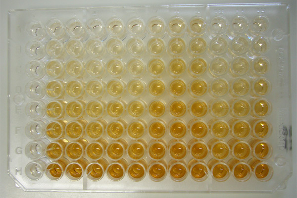


Basic principle and typical workflow
ELISA relies on two main components: a binding reagent (most often an antibody) that selectively captures the target molecule, and an enzyme label that produces a detectable signal proportional to the amount of target present. A typical workflow includes coating, blocking, sample incubation, detection with an enzyme-conjugated antibody, and signal development. Steps and reagent choices are optimized to minimize non-specific binding and maximize sensitivity.
- Coating: immobilize a capture antibody or antigen on a microplate well to retain the target.
- Blocking: apply an inert protein or blocking buffer to cover uncoated surfaces and reduce background.
- Sample incubation: add the sample so the target can bind to the immobilized capture reagent; wash away unbound material.
- Detection: add a detection antibody that binds the captured target; this antibody is conjugated to an enzyme or is detected by an enzyme-linked secondary antibody.
- Signal development: add an enzyme substrate that yields a colorimetric, fluorescent, or chemiluminescent product; measure the signal with appropriate instrumentation.
Common ELISA formats
- Direct ELISA: an enzyme-conjugated primary antibody detects the antigen directly; it is simple but offers limited signal amplification.
- Indirect ELISA: a primary antibody binds the antigen and an enzyme-linked secondary antibody recognizes the primary, increasing sensitivity.
- Sandwich ELISA: a capture antibody immobilizes the antigen and a second detection antibody binds a different epitope; this format is highly specific and suited for complex samples such as serum or tissue culture supernatant.
- Competitive ELISA: sample antigen competes with labeled antigen for limited binding sites; useful for small molecules or when only one antibody is available.
Detection chemistry and readouts
Common enzyme labels include horseradish peroxidase (HRP) and alkaline phosphatase (AP). Readouts may be colorimetric (measured as absorbance), fluorescent, or chemiluminescent. Choice of substrate and detection mode affects sensitivity, dynamic range and ease of quantification. Quantitative assays typically use a standard curve prepared with known concentrations of the analyte to convert signal intensity into concentration.
Controls, quantification and interpretation
Reliable ELISA results require appropriate controls: blanks to assess background, negative controls to check specificity, and positive controls to confirm assay performance. A series of known standards is used to construct a calibration curve for quantification. Matrix effects from complex samples can alter assay behavior, so samples are often diluted and spiked recovery tested. Assay linearity, limit of detection, and precision are commonly evaluated during validation.
Sample types and preparation
ELISA accepts a variety of sample types including serum, plasma, urine, saliva and culture supernatants. Proper sample handling—such as avoiding multiple freeze–thaw cycles, clarifying particulates, and using protease inhibitors when appropriate—helps preserve analyte integrity. Pre-dilution, centrifugation and use of compatible buffers can reduce interferences.
Sensitivity, specificity and limitations
ELISA is valued for good specificity when high-quality antibodies are used, but cross-reactivity can occur if antibodies recognize similar epitopes. Sensitivity depends on antibody affinity, enzyme substrate, and instrumentation. Limitations include potential matrix interferences, requirement for well-characterized reagents, and less suitability for multiplexing many analytes in a single well compared with bead-based platforms.
Derivatives, alternatives and related methods
Several methods build on ELISA principles. The ELISPOT technique adapts ELISA chemistry to detect molecules secreted by individual cells and to enumerate secreting cells; more on that approach is available at ELISPOT references. Multiplex immunoassays and bead-based platforms allow parallel measurement of multiple analytes. For specific applications, such as measuring small antigens or cytokine panels, consult method-specific resources like cytokine assay guides and antigen detection summaries.
Best practices and troubleshooting
Optimize blocking conditions, wash stringency and antibody concentrations to reduce background. Validate each antibody pair for sandwich assays to ensure they bind distinct epitopes. If signal is weak, consider increasing incubation times, improving enzyme activity or using more sensitive substrates. If background is high, evaluate blocking buffers, reduce antibody concentrations, or increase wash steps. Manufacturer protocols and reagent datasheets are useful starting points; for antibody sourcing and characterization consult antibody resources.
Applications and significance
ELISA remains a cornerstone technique across clinical and research laboratories. It is used for serology, hormone and cytokine measurements, detection of infectious agents, vaccine immunogenicity studies, and quality control in biologics manufacturing. Its balance of specificity, adaptability and relative simplicity makes it a practical tool for many workflows, though careful design and validation are essential for robust results.


History
The precursor of the ELISA was the radioimmunoassay since 1960. For an enzymatic detection, the direct coupling of proteins was necessary so that the reporter signal also only appears coupled with the specifically binding antibody. Chemical coupling of proteins was developed simultaneously by Stratis Avrameas and G. B. Pierce. The adsorption of proteins on surfaces had already been studied by Jerker Porath in 1966. The ELISA was developed simultaneously in 1971 by two research groups, including Peter Perlmann and Eva Engvall in Sweden.
Signal amplification
Instead of an enzyme-coupled detection antibody, the combination of an uncoupled detection antibody and an additional (third) secondary antibody (secondary because it is an antibody against antibody), to which an enzyme has been bound, can also be used for signal amplification (see Fig.). This requires another incubation and washing step. The buffer used is usually TBS-T buffer. Although more laborious, the use of a secondary antibody conjugate has the advantage that the costly production of many different enzyme-linked primary antibodies specific for only one antigen each can be circumvented. The secondary enzyme-linked antibodies used, which are polyclonal antibodies that can bind simultaneously to different epitopes in the constant region (Fc region) of all the primary antibodies of a species, can be used more widely and lead to signal amplification. In addition, secondary antibody-enzyme conjugates can be used for a variety of different immunoassays due to specificity for Fc regions of an antibody subtype from a species, making the secondary antibody a more cost-effective industrial mass-produced product. Further common signal amplification can be achieved by binding streptavidin or avidin conjugates to biotinylated detection antibodies in the final incubation step. Detection with (strept-)avidin-enzyme conjugates also result in signal amplification due to multiple biotinylations of the primary antibodies and the consequent binding of multiple reporter molecules.
Modern reporter systems partly allow higher sensitivities (e.g. immuno-PCR) or parallel determinations in one approach (multiplex) by using fluorescence or the polymerase chain reaction, but are not ELISAs in the strict sense.
Questions and answers
Q: What is ELISA?
A: ELISA stands for Enzyme Linked Immuno-Sorbent Assay, which is a technique used in biochemistry to detect the presence of a specific substance within a sample.
Q: How does ELISA work?
A: ELISA uses antibodies that attach themselves to the substance being tested, generating a specific color that indicates the amount of the substance present.
Q: What kind of substances can ELISA detect?
A: ELISA can detect specific cytokines or antigens within a sample.
Q: What does the amount of color generated by ELISA indicate?
A: The amount of color generated by ELISA indicates the amount of the substance being tested present in the sample.
Q: Is ELISA the only technique used to detect specific substances within a sample?
A: No, there is a more sophisticated and sensitive technique called the ELISPOT method derived from the ELISA technique.
Q: What is the role of the second set of antibodies used in ELISA?
A: The second set of antibodies in ELISA is used to capture the substance being tested, adhering to both the substance and the testing container to hold the substance in place.
Q: Does ELISA always generate color when a substance is detected?
A: Not always, sometimes the substance must be viewed under ultraviolet light for the antibodies to generate the color.
Related articles
Author
AlegsaOnline.com ELISA: Enzyme-Linked Immunosorbent Assay — principles, types, and uses Leandro Alegsa
URL: https://en.alegsaonline.com/art/30844