Electron crystallography: principles, history, and applications
Electron crystallography determines atomic arrangements in solids and biological assemblies using transmission electron microscopes; it complements X-ray methods and excels with thin or small crystals.
Overview
Electron crystallography is a structural technique that determines the arrangement of atoms in materials and biological assemblies by analysing the way an electron beam scatters from a specimen. Unlike methods that rely solely on X-ray scattering, electron crystallography exploits the strong interaction between electrons and matter, allowing structural studies of very thin specimens, two-dimensional crystals and tiny three-dimensional crystals that are unsuitable for conventional X-ray crystallography. Key experimental hardware is the transmission electron microscope (TEM), which produces both images and diffraction patterns used to recover atomic structure.
Image gallery
2 Images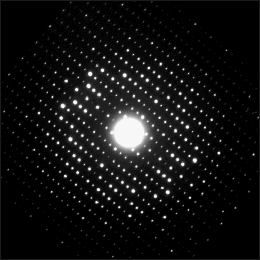

Principles and methodology
In practice, electron crystallography combines real-space imaging and reciprocal-space diffraction. High-resolution images capture projected densities while selected-area and convergent-beam diffraction give intensities for specific lattice planes. These signals are processed with Fourier methods to build three-dimensional maps. Careful control of sample thickness, orientation and dose is required because electrons interact strongly with atoms and multiple scattering can complicate intensity interpretation. Cryogenic sample preparation is commonly used to reduce radiation damage and preserve native states.
Sample types and common applications
The method is especially valuable for specimens that form regular, repeating arrangements but do not yield large single crystals. Typical targets include thin inorganic and organic solids, membrane proteins arranged in two-dimensional lattices, and symmetric biological assemblies. Protein studies often use 2D crystalline sheets or helical arrays (sheets or helices), polyhedral structures such as viral capsids (viral capsids), or highly ordered arrays. Electron crystallography is complementary to X-ray crystallography and other electron-based approaches and can reveal features that are inaccessible when large 3-D crystals are not available.
History and important milestones
The theoretical and practical foundations of electron crystallography were developed through mid-20th-century advances in electron microscopy and diffraction. The formalisation of crystallographic electron microscopy and its application to biological macromolecules is associated with Nobel-winning work by Aaron Klug. Landmark structural achievements include the high-resolution determination of membrane proteins and other macromolecular assemblies; one of the earliest widely cited protein structures solved by electron crystallography was bacteriorhodopsin, and subsequent studies reported structures such as light-harvesting complexes (light-harvesting complex) and the bacterial flagellum (bacterial flagellum).
Advantages, limitations and recent developments
- Advantages: can work with very small or thin crystals, provides both imaging and diffraction information, and is particularly suited to membrane proteins and virus particles.
- Limitations: strong electron–matter interaction leads to radiation damage and dynamical scattering; samples must be thin and well ordered; image interpretation requires careful computational correction.
- Recent advances: improved direct electron detectors, better image-processing algorithms, cryo-preparation techniques and electron diffraction variants (often used for tiny 3-D crystals) have expanded the technique's reach and resolution.
Workflow and notable distinctions
A typical workflow begins with specimen preparation to form well-ordered lattices or thin sections, data collection in a TEM to record images and diffraction patterns, and computational reconstruction that merges phase and amplitude information. Electron crystallography sits among other electron-based structural methods: it differs from single-particle cryo-EM in relying on lattice order, and from routine X-ray crystallography by enabling studies of systems that cannot be crystallised into large 3-D blocks. Practitioners often combine methods to validate models and to interpret functional details in materials and biological structures.
For conceptual background, instrument specifications and procedural details see references and resources linked to technical pages on atoms, solids, TEM, X-ray crystallography, 3-D crystals, proteins, helical arrays, polyhedrons, viruses and historical accounts of Aaron Klug.
Further technical examples and case studies can be explored through specialised literature and instrument manuals; detailed application notes often illustrate steps for membrane proteins, viral capsids and small-molecule crystals using electron crystallography protocols.
Questions and answers
Q: What is electron crystallography?
A: Electron crystallography is a method to determine the arrangement of atoms in solids using a transmission electron microscope (TEM).
Q: How does electron crystallography compare to X-ray crystallography?
A: Electron crystallography works in cases where X-ray crystallography does not. X-ray crystallography needs large 3-D crystals to work, while electron crystallography can be used with 2-dimensional crystals (sheets or helices), polyhedrons such as viral capsids, or dispersed proteins.
Q: Why can electrons be used in situations where X-rays cannot?
A: Electrons interact more strongly with atoms than X-rays do, which allows them to be used in situations where X-rays cannot.
Q: Who invented electron crystallography?
A: Aaron Klug invented electron crystallography.
Q: What did Aaron Klug win the Nobel Prize in Chemistry for?
A: Aaron Klug won the Nobel Prize in Chemistry for his invention of electron crystallography, as well as his studies on virus structures and transfer RNA.
Q: What was the first electron crystallographic protein structure to be solved?
A: The first electron crystallographic protein structure to be solved was bacteriorhodopsin in 1990.
Q: What are some other high-resolution structures that have been solved by electron crystallography?
A: Other high-resolution structures that have been solved by electron crystallography include the light-harvesting complex and the bacterial flagellum.
Related articles
Author
AlegsaOnline.com Electron crystallography: principles, history, and applications Leandro Alegsa
URL: https://en.alegsaonline.com/art/30732
Sources
- ncbi.nlm.nih.gov : PMID 8107845
- ncbi.nlm.nih.gov : PMID 12904785