Electrocardiography
![]()
ECG is a redirect to this article. For other meanings, see ECG (disambiguation).
The electrocardiogram (ECG) (from the ancient Greek καρδία kardía, German 'heart', and γράμμα grámma, German 'written') is the recording of the sum of the electrical activities of all the heart muscle fibers by means of an electrocardiograph (also called an ECG machine). The recording process is called electrocardiography. In German, the electrocardiogram is also known as the heart voltage curve, and is sometimes also called the heart script.
Every contraction of the heart muscle is preceded by an electrical excitation, which normally originates from the sinus node. It travels to the other cardiac muscle cells via the existing electrical conduction system consisting of specialised cardiac muscle cells. These electrical voltage changes at the heart can be measured at the body surface and recorded over time. The result is a recurring picture of the electrical action of the heart. The ECG can be used to make a variety of statements about the characteristics and health of the heart. It should be noted that the surface ECG only indicates the electrical activity of the heart muscle, but does not reflect the actual ejection fraction. In most cases, the ECG is evaluated by increasingly reliable computer programs, which, however, does not make the evaluation of the recording on paper or on the screen by the physician superfluous.

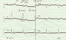
typical 6-channel ECG

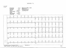
A normal 12-lead EKG...
History
In 1843, Carlo Matteucci discovered through experiments on pigeon hearts that cardiac activity is based on electrical processes. In 1882, physiologist Augustus Desiré Waller conducted the first ECG on his dog Jimmy by immersing its four paws in conductive sodium chloride solution. In 1887, he was able to record heart currents for the first time using a capillary electrometer.
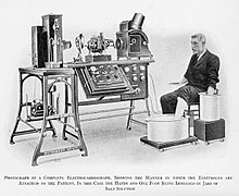
The instruments were substantially improved in 1903 by Willem Einthoven, who, building on his string galvanometer developed from 1895, developed the ECG into a useful diagnostic procedure and introduced it into the clinic. The terminology he introduced is still used today. He initially wanted to standardize on a single derivative in which the patient dips both arms into separate solutions (Einthoven I). Since this was not sufficient, the other extremity derivations Einthoven II (right arm - left leg) and III (left arm - left leg) were added, as well as later the Wilson derivations on the chest wall (after Frank Norman Wilson, 1934) and the Goldberger derivations (after Emanuel Goldberger, 1942), which are explained below.

Early form of ECG derivation according to Einthoven by immersion of the extremities in tubs with saline solution
Benefit
The ECG is a painless, non-invasive examination procedure that can be repeated at any time and performed almost anywhere.
The ECG can be used to determine the heart rate, heart rhythm and position type (electrical heart axis, cf. Cabrera circuit) and to read the electrical activity of the atria and ventricles. The ECG is just as indispensable for diagnosing cardiac arrhythmias such as extra beats (extrasystoles) and disturbances in the conduction and propagation of excitation (e.g. bundle branch block and AV block) as it is for detecting myocardial ischemia or a heart attack. Disturbances of the excitation regression (repolarization) can lead to so-called ventricular end part changes (changes of the ST segment or the T wave). The activity of a pacemaker presents itself as a very narrow, vertical line (spike).
The ECG may also provide evidence of thickening of the heart wall (hypertrophy of the myocardium), abnormal right or left heart strain, inflammation of the pericardium (pericarditis) or heart muscle (myocarditis), as well as electrolyte disturbances and adverse drug reactions.
With regard to most diagnoses, the ECG only provides indications and must not be assessed independently of the clinical picture (e.g. myocardial infarction, signs of hypertrophy, myocarditis). Only in the case of disturbances of cardiac rhythm or conduction can a clear diagnosis usually be made from the ECG alone.
Questions and Answers
Q: What is an electrocardiogram?
A: An electrocardiogram (ECG or EKG) is a record of the electrical activity of the heart over a period of time.
Q: Who invented the Electrocardiograph?
A: Willem Einthoven invented the Electrocardiograph.
Q: How does an electrocardiogram work?
A: An electrocardiogram works by attaching electrodes to the outer surface of the skin to record the electrical signals as they travel through the heart.
Q: What can doctors diagnose using an electrocardiogram?
A: Doctors can diagnose various heart conditions, measure the rate and regularity of heartbeats, the size and position of the chambers, the presence of any damage to the heart, and the effects of drugs or devices used to regulate the heart using an electrocardiogram.
Q: Is an electrocardiogram a painful test?
A: No, an electrocardiogram is a painless test.
Q: When are the results of an electrocardiogram reported?
A: The results of an electrocardiogram are likely to be reported the same day it is done.
Q: What does an electrocardiogram monitor?
A: An electrocardiogram monitors the heart by recording each beat of the heart triggered by an electrical impulse.
Search within the encyclopedia