
Dentin: structure, formation, and clinical significance
Dentin is the calcified tissue forming the bulk of a tooth beneath enamel and surrounding the pulp. This article explains its composition, development, types, function, and dental relevance.
Overview
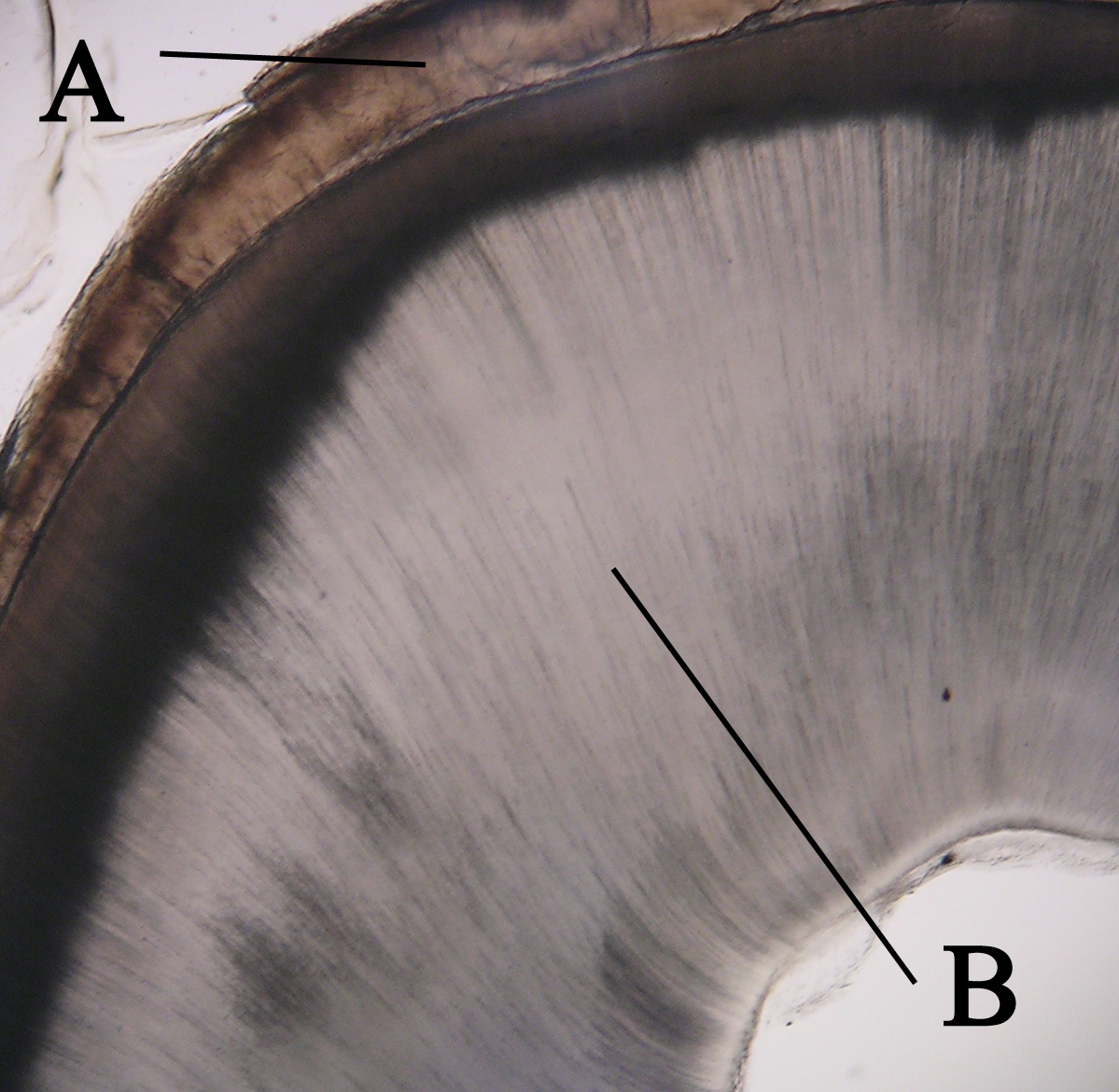
Dentin (or dentine) is the mineralized tissue that constitutes the main body of a tooth. In the crown it lies between the outer enamel and the inner dental pulp (pulp), and in the root it fills the space between the pulp and the thin outer cementum (cementum). Because enamel is semi‑transparent, the natural yellowish tint of dentin often affects overall tooth color (yellow hue).
Image gallery
4 Images

Structure and composition
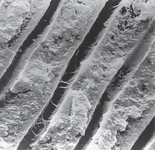
Dentin is softer and less mineralized than enamel but harder than bone. Microscopically it is built from a collagenous organic matrix that becomes impregnated with mineral crystals. Running through dentin are microscopic channels called dentinal tubules; each tubule contains a cellular process of an odontoblast, fluid, and nerve fibers that contribute to the tissue’s sensitivity.
- Odontoblasts: cells that form dentin and line the pulp chamber.
- Dentinal tubules: microscopic canals that alter permeability and conduct stimuli.
- Intertubular and peritubular dentin: matrix regions with different mineral densities.
Development and types
Dentin is formed by odontoblasts during tooth development in a process called dentinogenesis. Primary dentin forms as teeth develop, secondary dentin accumulates slowly after eruption throughout life, and tertiary (or reparative) dentin can be produced in response to injury, decay, or wear.
Function and clinical relevance
Dentin provides bulk and resilience to the tooth, absorbing forces that might otherwise crack enamel. Its tubules explain why exposed dentin produces sensitivity to temperature and touch. In dentistry, caries that penetrate enamel into dentin tend to progress faster; restorative and endodontic treatments focus on sealing, protecting, or removing affected dentin to preserve tooth function.
Notable distinctions and facts
Unlike enamel, dentin can continue to be deposited and altered throughout life. Age and reparative processes may darken dentin, affecting cosmetic appearance. Because it is living tissue connected to the pulp, dentin reacts biologically to stimuli and pathogens, making its preservation and proper restoration important in oral health care.
Questions and answers
Q: What is dentin?
A: Dentin is calcified tissue that makes up the second layer of teeth.
Q: Where is dentin found in the crown of a tooth?
A: In the crown of a tooth, dentin is found between enamel and pulp.
Q: Where is dentin found in the root of a tooth?
A: In the root of a tooth, dentin is found between cementum and pulp.
Q: What is the main component of a tooth?
A: Dentin makes up most of a tooth.
Q: What color is dentin?
A: Dentin is yellow.
Q: Why does the yellowness of dentin show through in a tooth?
A: The yellowness of dentin shows through in a tooth because enamel is semi-translucent.
Q: What are the layers of a tooth that dentin is found between?
A: In the crown of a tooth, dentin is found between enamel and pulp, while in the root of a tooth, it is found between cementum and pulp.
Related articles
Author
AlegsaOnline.com Dentin: structure, formation, and clinical significance Leandro Alegsa
URL: https://en.alegsaonline.com/art/26648
Sources
- commons.wikimedia.org : Dentin