Dendrite: structure, function, development, and biological significance
Dendrites are branched neuronal projections that receive and integrate synaptic input, enabling electrical signaling, plasticity, and complex computation across nervous systems.
Overview
Dendrites are the branched extensions of neurons that receive chemical and electrical signals from other cells and convey those inputs toward the cell body or soma. They form complex arborizations that vary widely in size, shape and branching pattern across neuron classes. Most neurons bear multiple dendrites but typically only a single axon, creating a polarized structure in which dendrites serve primarily for input and axons for output.
Image gallery
2 Images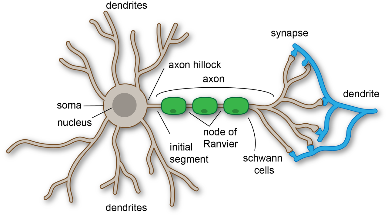

Structure and microanatomy
Dendrites are bounded by a specialized membrane rich in neurotransmitter receptors and voltage-gated ion channels. Many excitatory synapses form on small protrusions called dendritic spines; spines compartmentalize biochemical signals and can change shape rapidly. Other neurons have "smooth" dendrites without spines, commonly seen in many inhibitory interneurons. Inside, dendrites contain cytoskeletal elements (microtubules and actin), mitochondria and endoplasmic reticulum that support local metabolism, transport and signaling.
Synaptic input and electrical integration
Synaptic activation produces local changes in membrane potential: excitatory inputs depolarize the membrane, while inhibitory inputs hyperpolarize it. These postsynaptic potentials spread electrotonically along dendritic branches toward the soma. Temporal and spatial summation of many inputs determines whether the neuron reaches threshold for generating an action potential at the axon hillock. In addition to passive spread, dendrites express active conductances—voltage-gated sodium, calcium and potassium channels—that can amplify, shape or locally generate regenerative events such as dendritic spikes.
Computation and information processing
Dendrites contribute to neuronal computation beyond simple summation. Branches can perform local operations such as coincidence detection, subunit processing and nonlinear integration of inputs, effectively increasing the computational repertoire of single neurons. Backpropagating action potentials convey output-related signals into dendrites and interact with synaptic inputs to influence plasticity rules.
Development and plasticity
Dendritic arbors are shaped during development by genetic programs and extracellular cues. Synaptic activity refines branching and spine structure: long-term potentiation and long-term depression are associated with spine enlargement and shrinkage or elimination, respectively. Adult stem cells in restricted regions can generate new neurons that extend dendrites as they integrate into existing circuits. Throughout life, dendrites retain some capacity for structural remodeling in response to experience, learning and injury.
Roles in health and disease
Normal dendritic architecture and spine density are critical for cognitive function. Alterations in dendritic structure, spine morphology or synaptic receptor composition are observed in developmental disorders, psychiatric conditions and neurodegenerative diseases. Toxic insults, metabolic stress and genetic mutations can disrupt dendritic signaling, contributing to circuit dysfunction.
Experimental study and applications
Investigators study dendrites using electrophysiology (patch-clamp recordings from somata and dendrites), optical imaging (calcium and voltage indicators), and high-resolution microscopy to visualize spine dynamics and ultrastructure. Computational models of dendritic trees help relate morphology and channel distributions to neuronal input–output properties. Understanding dendritic function informs approaches to cognitive enhancement, neurorehabilitation and treatments for neurological disorders.
Summary and related topics
- Dendrites receive and integrate synaptic inputs and influence whether a neuron fires an action potential.
- Dendritic spines are key sites for excitatory synapses and for synapse-specific plasticity.
- Diversity in dendritic form and active properties supports varied signaling roles across neuron types.
For additional context, see entries on neurons, the soma, the axon, and adult neural stem cells for how new neurons develop dendritic arbors.
Questions and answers
Q: What are dendrites?
A: Dendrites are the branches of neurons that receive signals from other neurons.
Q: Where do signals go after entering dendrites?
A: Signals go into the cell body (or soma) after entering dendrites.
Q: How many axons can a cell have?
A: A cell may have only one axon.
Q: What do dendrites carry from other neurons into the soma?
A: Dendrites carry signals from other neurons into the soma.
Q: What is a synapse?
A: A synapse is a very narrow gap between the dendrite of one neuron and the axon of another neuron.
Q: What triggers the release of neurotransmitters?
A: Electrical impulses reaching the end of an axon trigger the release of neurotransmitters.
Q: What happens if dendrites get lots of signals from axons?
A: If dendrites get lots of signals from axons, it sets off a chain reaction called an action potential that flows down the axon to the next synapse.
Related articles
Author
AlegsaOnline.com Dendrite: structure, function, development, and biological significance Leandro Alegsa
URL: https://en.alegsaonline.com/art/26546
Sources
- faculty.washington.edu : "Brain Facts and Figures"
- doi.org : 10.1038/nrn2124
- pubmed.ncbi.nlm.nih.gov : 17453017
- books.google.com : Developmental biology