
Cranial nerve: structure, function, and clinical significance
Cranial nerves are paired nerves emerging from the brain or brainstem that carry sensory, motor and autonomic signals for the head, neck and some thoracic or abdominal structures.
Overview
A cranial nerve is any nerve directly attached to the brain or brainstem rather than to the spinal cord. Unlike spinal nerves, which emerge from segments of the spinal cord, cranial nerves provide more immediate channels between the central nervous system and structures of the head, neck and, in some cases, the thorax and abdomen. Each cranial nerve exists as a left and right pair and together they perform sensory, motor and autonomic roles that are essential for everyday functions such as smell, vision, facial movement, hearing and swallowing.
Image gallery
10 Images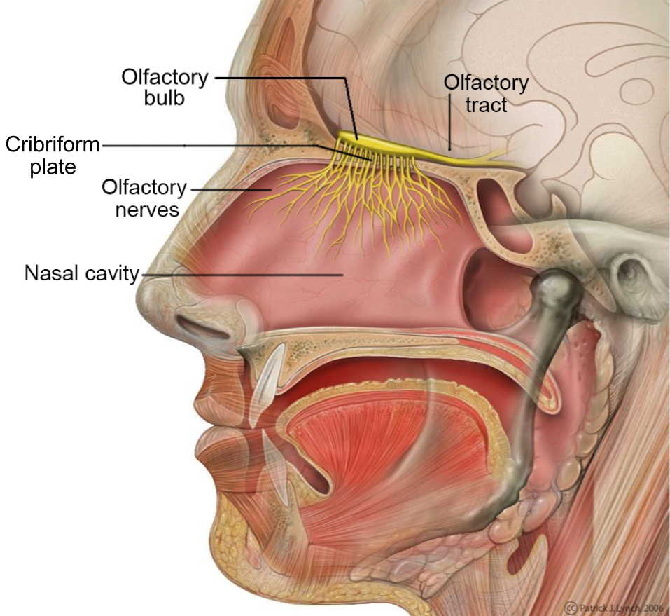



Names, numbers and basic functions
In humans there are twelve pairs of cranial nerves. They are traditionally numbered from front to back with Roman numerals I–XII. Each nerve has a name that reflects its principal function or distribution. The sequence and general modalities are widely taught and used in clinical practice:
- I Olfactory — smell (sensory)
- II Optic — vision (sensory)
- III Oculomotor — most eye movements and pupil control (motor/autonomic)
- IV Trochlear — a single extraocular muscle (motor)
- V Trigeminal — facial sensation and mastication (mixed)
- VI Abducens — lateral eye movement (motor)
- VII Facial — facial expression, taste anterior tongue, lacrimation and salivation (mixed)
- VIII Vestibulocochlear — hearing and balance (sensory)
- IX Glossopharyngeal — taste posterior tongue, swallowing, salivation (mixed)
- X Vagus — parasympathetic control to thoracic and abdominal organs, voice and swallowing (mixed/visceral)
- XI Accessory — sternocleidomastoid and trapezius muscles (motor)
- XII Hypoglossal — tongue movement (motor)
Anatomy and development
Each cranial nerve arises from specific nuclei or rootlets within the brain or brainstem and exits the skull through foramina to reach its target tissues. The anatomical relationships are complex: some fibers originate from central neurons, others from peripheral sensory ganglia whose cells derive in part from neural crest or ectodermal placodes. All cranial nerves are found above the level of the first vertebra in the neck, and they traverse regional structures of the head and neck to reach muscles, glands and sensory receptors. The sensory and motor components are organized according to modality—somatic, visceral, special senses and branchial motor (also called special visceral efferent).
History and terminology
The classification and numbering of cranial nerves evolved as anatomists described their origins and functions. The paired arrangement and the front-to-back numbering convention are long established in anatomy texts. Historical debates have concerned the exact contributions of some nerves (for example, the accessory nerve's spinal rootlets) and the terminology for special fibers; modern descriptions integrate macroscopic anatomy with embryology and physiological testing.
Clinical relevance and testing
Cranial nerves are routinely examined during a neurological assessment because their function reflects specific brainstem and forebrain pathways. Deficits can present as loss of smell or vision, double vision, facial weakness, altered hearing or balance, difficulties swallowing or changes in voice. Simple bedside tests evaluate individual nerves: smell tests for I, visual acuity and fields for II, ocular movements for III–VI, facial sensation and power for V and VII, hearing and balance tests for VIII, and gag, phonation and shoulder shrug for IX, X and XI. Disorders affecting cranial nerves range from infections and inflammatory conditions to traumatic injury, ischemia and tumors.
Distinctions and notable facts
Cranial nerves differ from spinal nerves in distribution and function: most spinal nerves serve the trunk and limbs, while cranial nerves predominantly serve the head and neck with a few exceptions such as the vagus nerve extending into the chest and abdomen. Each cranial nerve pair contributes discrete and often testable abilities, making them invaluable for localizing neurological lesions within the brain or brainstem. For further anatomical diagrams, clinical protocols and teaching resources see authoritative references and educational materials (brain anatomy overview, central nervous system resources, cranial nerve definitions, skull base foramina, neck anatomy, spinal vs cranial nerves, nerve numbering and names).
Questions and answers
Q: What is a cranial nerve?
A: A cranial nerve is any nerve that is directly attached to the brain or brainstem.
Q: How does the attachment of cranial nerves differ from spinal nerves?
A: Spinal nerves are attached to segments of the spinal cord, whereas cranial nerves are attached directly to the brain or brainstem.
Q: Which parts of the body do cranial nerves mostly relay information between?
A: Cranial nerves mostly relay information between parts of the head and neck.
Q: How many cranial nerves pairs are present in humans?
A: In humans, there are twelve cranial nerves pairs.
Q: Where do all cranial nerves appear in the body?
A: All cranial nerves appear above the first vertebra in the neck (cervical vertebra).
Q: How are cranial nerves numbered?
A: Cranial nerves are numbered using Roman numerals I–XII based on their order from the front of the brain to the back, where the brainstem is.
Q: Are cranial nerves present on both sides of the central nervous system?
A: Yes, each cranial nerve exists as a pair and is present on both sides of the central nervous system.
Related articles
Author
AlegsaOnline.com Cranial nerve: structure, function, and clinical significance Leandro Alegsa
URL: https://en.alegsaonline.com/art/23957
Sources
- accessmedicine.mhmedical.com : Clinical Neuroanatomy, 28e
- medicine.yale.edu : "Olfactory Nerve > Cranial Nerves | Yale School of Medicine"
- ncbi.nlm.nih.gov : "Regeneration of olfactory receptor cells"
- worldcat.org : 0300-5208
- pubmed.ncbi.nlm.nih.gov : 1752165
- books.google.com : Your Inner Fish: The amazing discovery of our 375-million-year-old ancestor