Corpus cavernosum clitoridis
Paired erectile bodies within the clitoris composed of spongy tissue that engorge with blood during arousal; homologous to the male corpora cavernosa and linked to sexual function and pelvic anatomy.
Overview
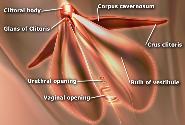
The corpus cavernosum clitoridis refers to one of two paired masses of erectile, spongy tissue that make up the internal body of the clitoris. Each corpus expands with blood during sexual arousal and contributes to clitoral rigidity and sensitivity. These structures are the female counterpart of the male corpora cavernosa and share a common embryological origin.
Image gallery
3 Images

Anatomy and relationships
Each corpus cavernosum lies along the crura of the clitoris and extends from the body toward the pubic arch. Externally the clitoral glans overlies the distal tips of these erectile bodies. Unlike the male anatomy, females do not have a distinct corpus spongiosum; instead, the vestibular bulbs lie alongside the vaginal entrance and expand in coordination with the corpora cavernosa. The erectile tissue is typically described as spongy and is a key component of the clitoris.
Vasculature, innervation, and function
Engorgement of the corpora results from increased arterial inflow and restricted venous outflow. They receive blood from branches of the internal pudendal artery and are connected to surrounding blood vessels. Sensation is carried chiefly by the dorsal nerve of the clitoris. Functionally, the corpora cavernosa contribute to sexual arousal, tactile sensitivity, and cushioning of the glans during sexual activity; the nearby vestibular bulbs also enlarge and may assist in shaping the vestibule at the entrance to the vagina.
Development and homology
Embryologically, the corpora cavernosa of the clitoris develop from the same tissues that form the male corpus cavernosum penis. Their growth and differentiation are influenced by hormonal environment during fetal development. In adults they remain homologous structures, adapted to female pelvic anatomy and reproductive physiology.
Clinical and notable points
- Because of their role in sexual function, the corpora cavernosa are considered in discussions of sexual health, clitoral surgery, and genital reconstruction.
- Damage, scarring, or surgical alteration can affect sensitivity and erectile capacity; preservation of vascular and neural connections is important during procedures.
- Anatomical descriptions often reference attachment to the pubic arch and nearby pelvic bones; some descriptions note connections to the pubic bone and hip bone region.
For imaging and research, techniques such as ultrasound and magnetic resonance have been used to visualize clitoral erectile tissues and their blood flow, helping clinicians and researchers better understand normal variation and functional changes.
Questions and answers
Q: What is the corpus cavernosum clitoridis?
A: The corpus cavernosum clitoridis is one of two sponge-like tissue found in the body of the clitoris.
Q: How is the corpus cavernosum clitoridis similar to the corpus cavernosum penis?
A: The corpus cavernosum clitoridis is similar to the corpus cavernosum penis in the male.
Q: Does the female have a corpus spongiosum?
A: No, the female has no corpus spongiosum.
Q: What does the female have instead of a corpus spongiosum?
A: The female has two vestibular bulbs beneath the skin at the entrance to the vagina, which expand at the same time as the glans clitoris.
Q: How is each corpus connected to the blood vessels of the pubic bone and hip bone?
A: Each corpus is connected to the blood vessels of the pubic bone and hip bone by the clitoris.
Q: What is the function of the corpus cavernosum clitoridis?
A: The function of the corpus cavernosum clitoridis is to become engorged with blood during sexual arousal, causing the clitoris to become enlarged and stiff.
Q: What is the significance of the vestibular bulbs?
A: The vestibular bulbs play an important role in female sexual response by expanding during arousal, contributing to the sensation of tightness and the increase in size of the vagina.
Author
AlegsaOnline.com Corpus cavernosum clitoridis Leandro Alegsa
URL: https://en.alegsaonline.com/art/23220