Corpus callosum: structure, function, development and clinical significance
The corpus callosum is the largest cerebral commissure in placental mammals, linking left and right hemispheres to enable interhemispheric communication, coordination, and transfer of information.
Overview
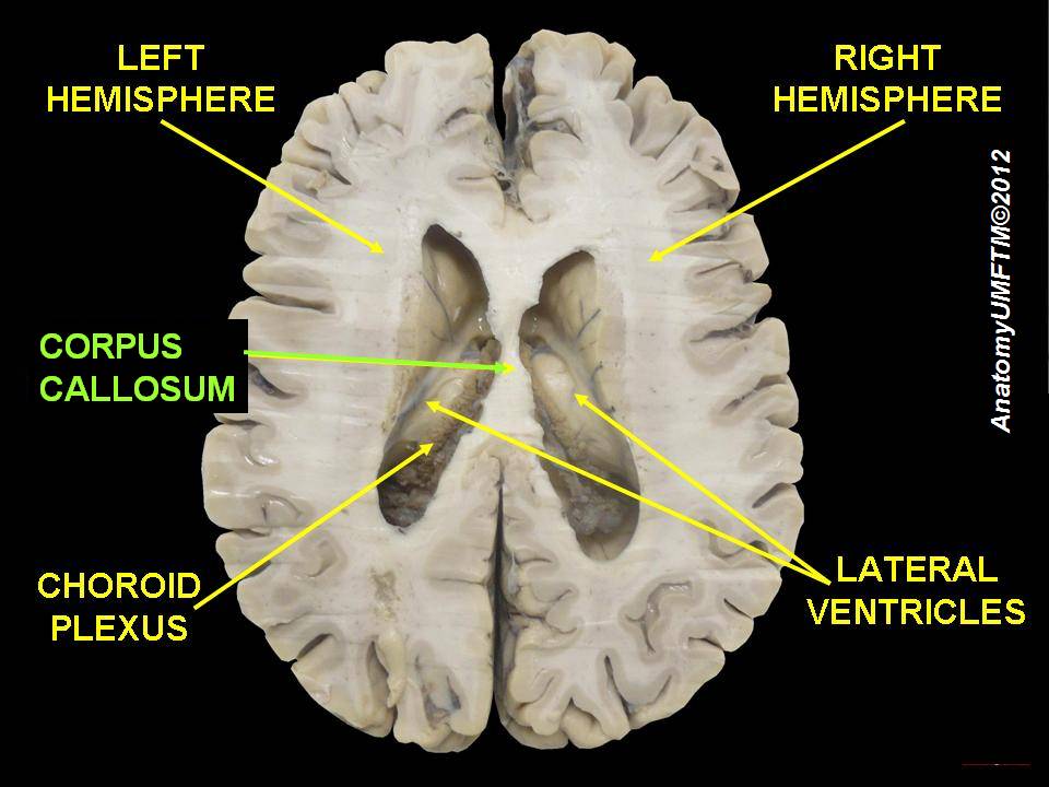
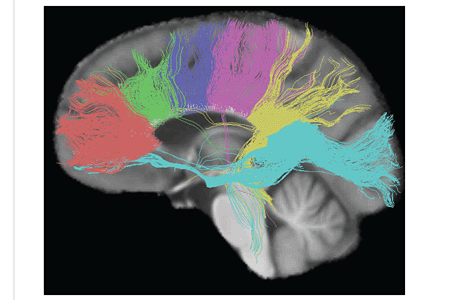
The corpus callosum is a broad bundle of nerve fibers that connects the left and right cerebral hemispheres in placental (eutherian) mammals. It is the principal pathway for interhemispheric communication, composed largely of myelinated axons. Estimates commonly describe it as containing over 200 million nerve fibers, though counts vary by species and methodology. By enabling signals to cross between hemispheres, the corpus callosum supports integrated perception, coordinated movement, and the sharing of cognitive processes.
Image gallery
10 Images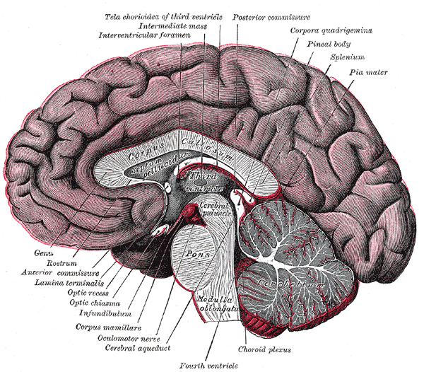


Anatomical parts and characteristics
Observed in midsagittal sections, the corpus callosum is conventionally divided into regions that differ in fiber composition and cortical connections. From front to back these regions are often named:
- Rostrum — anteroinferior portion connecting frontal polar regions.
- Genu — anterior bend linking prefrontal areas.
- Body (trunk) — central portion conveying fibers for motor and somatosensory cortices.
- Isthmus — a narrow region toward the posterior.
- Splenium — posterior thickened segment connecting parietal, temporal and occipital cortices.
Development and evolution
The corpus callosum forms during fetal and early postnatal development as axons cross the midline guided by molecular cues and transient cell populations. It is a feature of placental mammals; other vertebrates and non-placental mammals use different commissural systems, such as a relatively larger anterior commissure, for interhemispheric transfer. Abnormal development can result in partial or complete agenesis, with a wide range of functional outcomes.
Functions and importance
Functionally, the corpus callosum facilitates the exchange and integration of sensory, motor and higher-order information between hemispheres. Its role is central to coordinated bilateral movement, unified visual perception, and the transfer of learned information. It interacts with cortical specialization and lateralization of brain function, permitting hemispheric specialization while preserving cooperation.
Clinical relevance and notable facts
Damage, congenital absence, demyelinating disease, or surgical sectioning (callosotomy) affect interhemispheric communication and can produce characteristic signs, such as difficulties in tasks that require transfer of information between visual fields. Imaging methods like MRI and diffusion techniques are used to visualize corpus callosum anatomy and integrity. Some aspects such as sex differences in size or function remain subjects of ongoing research and debate.
Summary
As the brain's largest white-matter commissure, the corpus callosum is essential for integrating the two cerebral hemispheres. Its structure, development, and vulnerability to disease make it a frequent focus in neuroscience, neurology, and neuroimaging research.
Questions and answers
Q: What is the corpus callosum?
A: The corpus callosum is a part of the brain in humans and other eutherian mammals.
Q: What is the function of the corpus callosum?
A: The function of the corpus callosum is to connect the left and right hemispheres of the brain through over 200 million nerve fibers.
Q: Why is the corpus callosum important?
A: The corpus callosum is important because it relates to the lateralization of brain function, allowing the two sides of the brain to specialize in doing somewhat different things.
Q: What is lateralization of brain function?
A: Lateralization of brain function is where the two sides of the brain specialize in doing somewhat different things.
Q: How many nerve fibers connect the left and right hemispheres of the brain?
A: Over 200 million nerve fibers connect the left and right hemispheres of the brain.
Q: What is the largest connective pathway in the brain?
A: The corpus callosum is the largest connective pathway in the brain.
Q: Do all mammals have a corpus callosum?
A: No, only humans and other eutherian mammals have a corpus callosum.
Related articles
Author
AlegsaOnline.com Corpus callosum: structure, function, development and clinical significance Leandro Alegsa
URL: https://en.alegsaonline.com/art/23219
Sources
- commons.wikimedia.org : Category:Corpus callosum
- ncbi.nlm.nih.gov : ncbi.nlm.nih.gov/pmc/articles/PMC2770441/
- ncbi.nlm.nih.gov : "Absence of the corpus callosum as a mendelizing character in the house mouse"
- ui.adsabs.harvard.edu : 1933PNAS...19..609K
- doi.org : 10.1073/pnas.19.6.609
- jstor.org : 86284
- pubmed.ncbi.nlm.nih.gov : 16587795
- books.google.com : p. 68
- books.google.com : p. 50
- books.google.com : p. 175
- books.google.com : p. 361