Comparative anatomy: structure, methods, history, and applications
Comparative anatomy compares animal form and structure to infer function, development and relationships, using dissection, microscopy, collections and modern imaging and molecular methods for research and education.
Comparative anatomy is the systematic study of similarities and differences in the bodies of animals, used to infer how structures work, how they develop, and how they relate by descent. The field examines organs, tissues and skeletal elements across species to identify patterns of sameness and difference. For general discussion of animal form see animal bodies, and for more on internal organization see internal anatomy. Large-scale classification often reflects anatomical features; for an overview of higher groups consult the List of animal phyla.
Image gallery
5 Images


Core concepts
Two central ideas in comparative anatomy are homology and analogy. Homologous structures are similar because of common ancestry, while analogous structures perform similar functions but evolved independently. Distinguishing homology from convergence or homoplasy is essential for reconstructing evolutionary history and for avoiding misclassification. Comparative anatomical study therefore informs functional interpretation: for example, forelimbs modified as a wing, flipper or hand can be homologous yet have different mechanical roles.
Methods and sources of evidence
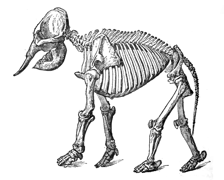
Historically, dissection has been the primary way to reveal organ relationships. Dissection remains a core educational and research technique, rooted in an ancient practice of opening specimens to study their internal structure. Microscopy expanded anatomical resolution: light and electron microscopy reveal cells and tissues, and modern histology links microscopic pattern to function. Collections housed in natural history museums and osteological repositories provide comparative material across time and geography for hypothesis testing.
- Specimen techniques: careful dissection, skeletal preparation and fixation are indispensable for revealing gross anatomy and muscle attachments.
- Microscopy and histology: tissue-level studies clarify organ architecture and developmental pathways; molecular markers such as DNA can be used to relate genetic variation to structural differences.
- Imaging and digital methods: non-destructive imaging (CT, MRI, micro-CT) and 3D reconstruction allow internal anatomy to be visualized in situ and compared quantitatively.
- Quantitative approaches: geometric morphometrics, biomechanical modelling and statistical comparison improve objectivity when comparing shapes and functions.
History and scientific role
The discipline matured from the late 18th through the mid-20th centuries and played a central role in debates about classification and origin. Anatomists and naturalists such as Georges Cuvier emphasized functional integration and the fossil record, while others like Thomas Henry Huxley used comparative data to argue for common descent. Charles Darwin relied on anatomical comparisons in several studies, notably his work on barnacles, to document variation and homologies. Some early practitioners opposed evolutionary interpretation; later work integrated anatomy with genetics and the fossil record to form modern evolutionary synthesis.
In contemporary biology, comparative anatomy complements molecular and genetic approaches such as molecular evolution and sequence-based phylogenetics using sequence analysis. Molecules provide an independent line of evidence for relationships, while anatomy provides the phenotypic context that links genes to functional form. For many research programs, anatomical observation remains indispensable: field biologists and zoologists use dissections and collections in ecological and taxonomic studies, and paleontologists infer soft tissues from bones and traces.
Educational and practical applications
Hands-on anatomical study is required in medical and veterinary training because understanding the human body and other organisms depends on direct knowledge of structural relationships. Comparative anatomy underpins taxonomy, functional morphology, and conservation biology. Professional research in these areas often relies on combining anatomical data with ecological, developmental and molecular evidence. A university degree in biology typically includes anatomy and morphology as foundational subjects, and anatomically based methods extend to plant structure, where comparative study of plants informs botany and paleobotany.
Interpretation and limitations
Interpreting anatomical similarities requires careful attention to developmental origin and function. Embryological data can reveal whether structures share a common developmental pathway, strengthening a claim of homology. Fossils extend comparisons into deep time but often preserve only hard parts; here, careful anatomical inference is needed to reconstruct muscles and organs. While molecular data are powerful, they do not replace the need to understand morphology: anatomy links genotype to phenotype and provides the observable traits by which organisms interact with environments.
In sum, comparative anatomy is a multidisciplinary enterprise that integrates dissection, microscopy, collections-based study, imaging and genetic approaches to explain organismal form and history. Readers seeking more on museum collections or modern analytical techniques may consult resources on natural history curation and digital imaging in comparative research (museum collections and molecular evolution resources). For discussion of broader evolutionary concepts see materials about evolution and its history.
Further reading and practical guides often cover core techniques and case studies that illustrate how anatomical evidence is gathered and interpreted. Historical accounts highlight contributions by figures such as Cuvier and Huxley, while modern reviews emphasize integrative approaches that combine anatomy with genetic and developmental data. Whether for teaching, taxonomy, paleontology or functional analysis, comparative anatomy remains a central discipline for understanding life’s form and diversity.
Questions and answers
Q: What is comparative anatomy?
A: Comparative anatomy is the scientific comparison of animal bodies in order to understand their working structure and determine the phylogenetic relationships between different groups of animals.
Q: What techniques are used for comparative anatomy?
A: The main techniques used for comparative anatomy are dissection and microscopy. Dissection involves examining the inside structure of a living thing, usually after it has died, while microscopy uses simple or compound microscopes to view small details of structure. Additionally, careful comparison of large collections of animals (usually in museums) is often done.
Q: When was the great era of comparative anatomy?
A: The great era of comparative anatomy was from about 1800 to about 1950.
Q: Who used comparative anatomy during this time period?
A: During this time period, both those who did not believe in evolution such as Georges Cuvier and those who did such as Thomas Henry Huxley used comparative anatomy. Charles Darwin himself also used it as his main tool when researching barnacles.
Q: What method is now mainly used to find out relationships between animals?
A: Molecular evolution is now mainly used to find out relationships between animals, which uses DNA sequence analysis.
Q: Is dissection still being done by zoologists today?
A: Yes, dissection is still being done by zoologists today for many research purposes.
Q: Do you need to know about the structure of animals and plants in order to get a degree in biology?
A: Yes, knowledge about the structure of animals and plants is necessary in order to get a degree in biology.
Related articles
Author
AlegsaOnline.com Comparative anatomy: structure, methods, history, and applications Leandro Alegsa
URL: https://en.alegsaonline.com/art/22215