Albinism: causes, characteristics, and impacts in humans, animals, and plants
Albinism is a congenital condition marked by reduced or absent melanin production, affecting skin, hair and vision. This article explains causes, types, health issues, and effects on animals and plants.
Albinism is a group of inherited conditions defined by reduced or absent production of the dark pigment known as pigment in cells. The most familiar pigment affected is melanin. People born with albinism may be described as albinos, though many prefer the term "person with albinism." The condition occurs in humans, various animals and even in plants, and leads to a recognizable range of physical and visual traits.
Image gallery
10 Images


Typical characteristics and underlying cause
Albinism commonly alters the color of hair, eyes and skin, producing very light or white hair, pale skin and eye colors that range from blue to gray; in some instances the eyes can appear pinkish because of visible blood vessels in the absence of pigment. The root cause is genetic: mutations in genes that control melanin synthesis or its distribution. These mutations reduce or block the activity of enzymes and pathways that manufacture melanin.
Vision is frequently affected because melanin contributes to normal eye development. Common visual issues include:
- Reduced visual acuity and poor depth perception.
- Nystagmus (uncontrolled eye movements) and strabismus (misaligned eyes).
- Photophobia (sensitivity to bright light) and foveal underdevelopment, which impair fine central vision.
Types and how common it is
Medical classification groups albinism into main categories: oculocutaneous albinism (affecting skin, hair and eyes) and ocular albinism (primarily affecting the eyes). Within these categories there are several genetic subtypes, often designated by laboratory and clinical names (for example OCA types and ocular forms), and some syndromes include albinism as one feature. Prevalence varies by population; albinism is rare overall, occurring at approximately a few dozen to a few thousand individuals per million depending on region and ancestry.
Health implications and daily life
People with albinism face two broad health concerns. First, the lack of skin pigment increases vulnerability to ultraviolet radiation: affected individuals can become sunburnt easily and have a higher lifetime risk of skin damage and skin cancers unless protected. Second, visual impairment can affect education, mobility and employment; many benefit from optical aids, tinted lenses, low-vision services and specialized teaching strategies.
Beyond medical matters, people with albinism often confront social and cultural challenges. Myths and misunderstanding can lead to stigma, discrimination, or unsafe situations in some societies. Advocacy groups and clinical services emphasize respectful language, inclusive education, and access to sun protection and eye care.
Albinism in animals and plants
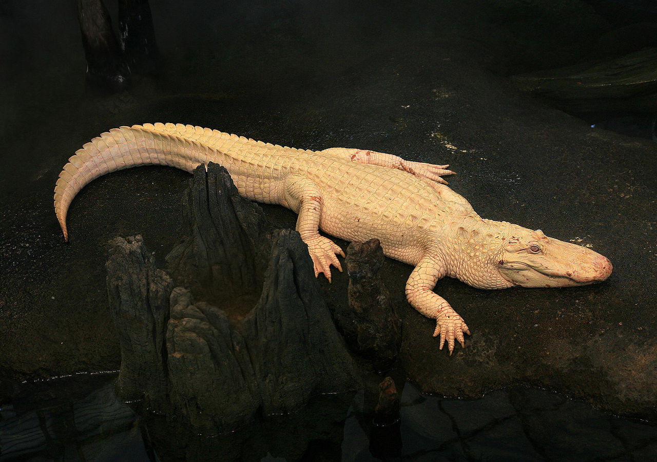
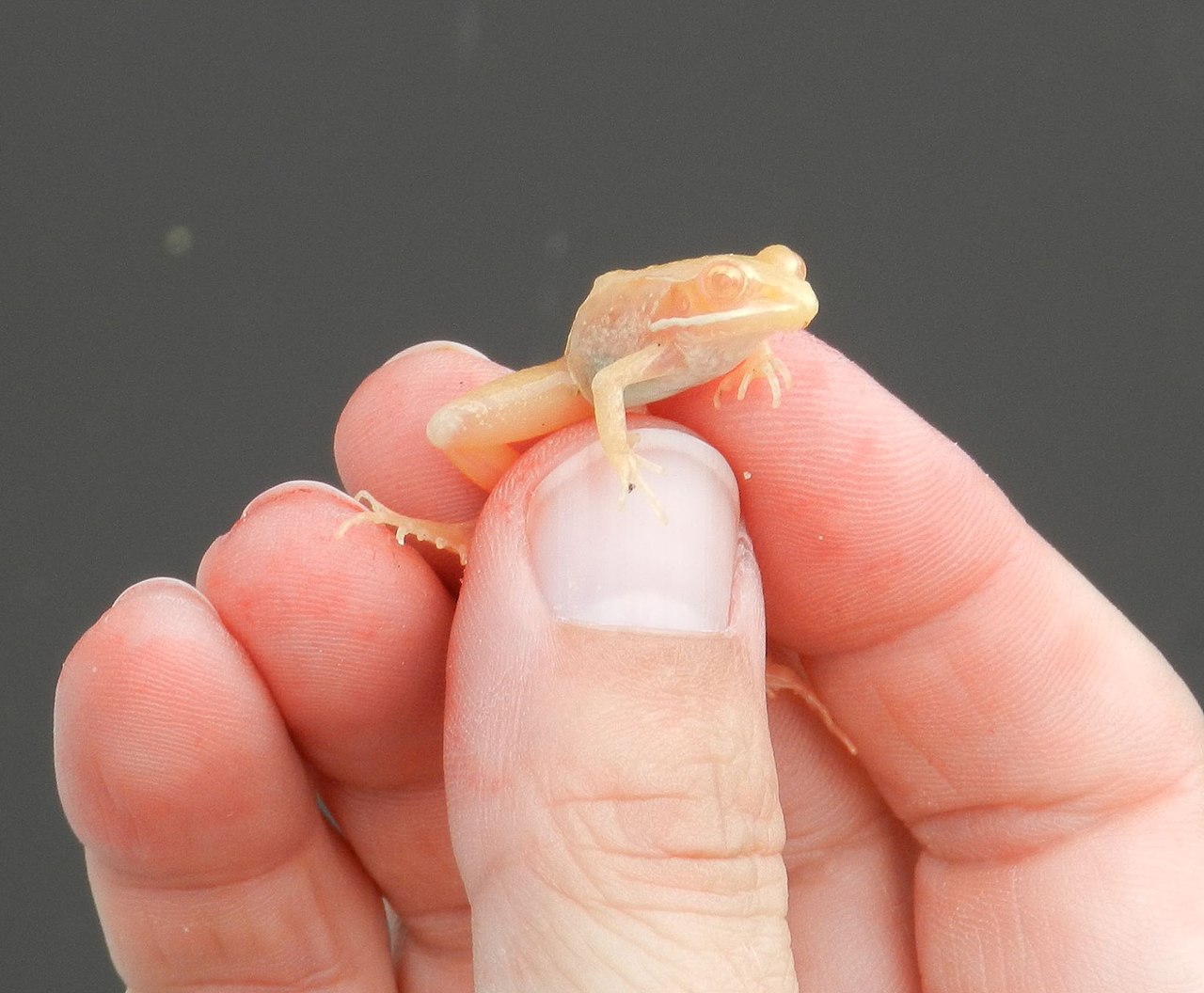
In wildlife and domestic species, albinism influences survival and behavior. Without normal coloration, animals may be conspicuous to predators, lack effective camouflage, or be disadvantaged in mate selection. Captive and laboratory animals are sometimes studied to learn how pigment genes work, but in the wild albino individuals are often rare. In the plant world, pigment loss affects chlorophyll or similar pigments; truly albino plant tissues cannot perform normal photosynthesis and often fail to survive past seedling stages.
Understanding albinism requires both genetic and social perspectives: its biology explains the visible signs and health needs, while cultural awareness supports the rights and daily safety of people with the condition. For medical guidance, educational supports, and community resources, consult specialized clinics and advocacy organizations (see links and resources represented here: melanin overview, hair changes, eye effects, skin care, hair color variations, animal cases, plant examples, pigment biology, sun protection, eye movement disorders, camouflage and ecology).


Albinism in humans
Appearance and symptoms
Even people whose bodies cannot produce any melanin at all, i.e. who are completely albinotic, are not extremely noticeable in Central and Northern Europe, since here skin, hair and eyes lightened by partial albinism are the normal case as an adaptation to the lower solar radiation. Full albinism in humans leads to pink skin, white-blond hair and pink-blue eyes. People with weaker albinism are not always clearly identifiable as such by their appearance. They look lighter than family members without albinism, but usually a residual function of melanin production is still preserved, so that there are also dark-skinned people with albinism who have clearly brown skin and light brown eyes.
While most people with albinism have lighter eye and hair color than their non-albinotic blood relatives (oculocutaneous albinism, OCA), there are also cases of albinism in which the symptomatology is limited to eye damage alone, while they look normal on the outside (ocular albinism, OA).
Skin color
People with albinism have lightened skin. They get sunburn more easily and thus have a higher risk of skin cancer. The skin color of whites is lightened as an adaptation to the lower sunlight outside the tropics, among other things, by mutations of the albinism genes, but not to the extent that this causes recognizable eye damage.
Symptoms affecting vision
In pure ocular albinism and in all forms of complete or almost complete oculocutaneous albinism, a pronounced symptom complex is present in the eyes. However, color perception is normal because albinism has no effect on the formation of rhodopsin.
Lightening of the eye color
Human eye colors of the iris can vary from dark brown to light brown and green to gray and blue, or if the iris is pigmentless, appear red as the blood red vessels show through.
enlarge and show information about the picture
![]()
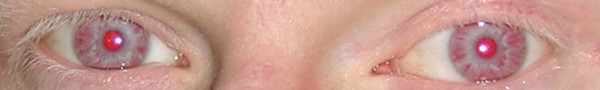
Eyes of a human with complete albinism (OCA1)
Albinism causes a lighter eye color. Complete albinism, regardless of what eye color the person would have had without their albinism, results in light blue, almost pink eyes as seen in the picture above, but this is very rare in humans. If very little melanin is produced, the eyes are blue. If there is a weaker albinism, where the body can still produce noticeable amounts of melanin, the eyes are correspondingly less brightly colored.
Light sensitivity
If the body can produce hardly any or almost no melanin and it is therefore not present in the eye or only to a very small extent, the iris becomes transparent to a certain extent and can be transilluminated with appropriate light. In less pronounced cases, the pigment defects are then found more in the area of the iris root. This transparency can also be seen by red light reflections when shining into the eye. Typical for people with high-grade albinism is therefore a pronounced sensitivity to glare (photophobia), which is why they usually wear sunglasses outdoors.
Disturbances of the spatial vision
Melanin also plays a role in the development of the optic nerves. Normally, the visual field in humans is divided equally between the two hemispheres of the brain - each hemisphere has its side and receives from both eyes the part of the image that belongs to that side (visual pathway). By comparing the two images, each hemisphere of the brain can calculate the distance of objects and assign them spatially (stereoscopic vision). In people with albinism, a larger proportion of the optic nerves cross to the opposite hemisphere of the brain, resulting in a loss of physiological adjacency of homologous retinal areas, and related images are not always processed on the same side.
In addition, ocular nystagmus (eye tremor) of varying severity is usually present, often accompanied by manifest strabismus (squint). In a series of examinations of 37 patients, all had nystagmus of various degrees, only four of them not squinting. The above-mentioned conditions therefore often result in absent or at least significantly limited spatial vision.
Decreased visual acuity
The visual fossa (fovea centralis), the retinal site of the sharpest vision, is anatomically not fully developed in albinism, as its development is also influenced by melanin. It is either not formed (aplasia) or develops only incompletely (hypoplasia).
Due to the nystagmus and the organic situation, the visual acuity in full-blown albinism is rarely more than 0.1, rather less, and can reach up to about 0.5 if the anomaly is less severe. Visual impairment varies, sometimes widely, even within the same type. Due to the existing pigment defects of the iris, contrasts between light and dark areas in space are often only indistinctly recognizable.
In addition, people with albinism are often unable to focus the eye at different distances (accommodation), and many sufferers are nearsighted or farsighted.
Hearing related symptoms
Very rare is the albinism deafness syndrome.
Treatment
Albinism does not affect the mental development of people. Therefore, despite the non-treatable metabolic defect, they can usually lead a largely normal life with the help of (magnifying) vision aids, tinted glasses or contact lenses and appropriate skin protection.
Physiology of albinism
Melanin synthesis
The pigment melanin is produced by pigment-forming cells, the melanocytes. The precursors of the melanocytes of the embryo, the melanoblasts, migrate during pregnancy in the early fetal period from the neural crest into the epidermis of the skin, into the hair follicles and various other organs. Once in the skin, the melanoblasts differentiate into melanocytes and form numerous cell processes through which they pass melanin to the keratinocytes. The amount of melanocytes in blacks is the same as in whites, and a person suffering from albinism also has a normal amount of melanocytes. Skin color is determined by the quantity and quality of the pigment melanin produced, not by the number of these cells.
Melanocytes contain melanosomes, small membrane-enclosed vesicles in which the pigment melanin is produced. They are very similar in function to lysosomes (cell organelles that serve digestion within the cell) because both contain substances that are dangerous to the cell and therefore must not come into contact with the rest of the cell. The lysosomes contain protein-dissolving enzymes (proteases) and the melanosomes contain intermediate products of melanin synthesis such as quinones and phenols, which can damage the cell's membranes.
In order to produce melanin, various enzymes are needed, which successively participate in the formation of melanin (gene action chain). If one of the enzymes of this metabolic pathway is no longer functional, albinism occurs. Eumelanin formation in melanosomes begins with hydroxylation of the amino acid (AS) L-tyrosine by the membrane-bound enzyme tyrosinase. In addition to this key enzyme, two other enzymes, DHICA oxidase and Dct, which are also membrane-bound, are required for eumelanin to be formed.
Molecular genetic classification of albinism
Although differences in the appearance of people with albinism were described early on, it was assumed that albinism was due to changes in a single gene. It was not until the family described by Trevor-Roper in 1952, in which both parents were affected by albinism and yet had normally pigmented children, that the first indication of the genetic heterogeneity of this disorder was given. Both parents in this case were homozygous (homozygous) for gene mutations leading to albinism. However, these affected different genes, so that the children were heterozygous (heterozygous) for each of the two mutations and thus were not clinically affected by albinism.
At first, albinism was classified according to external appearance. Later, it was possible to prove whether tyrosinase - an enzyme necessary for melanin production - was present. With the possibility of identifying some of the genes responsible for OCA, a molecular genetic classification was finally established. It was found that the different phenotypes are not always due to mutations in different genes, but often represent different expressions of diverse mutations in one gene. Clinical differentiation remains difficult, as it is not possible to infer the causative mutation (genotype) from the appearance (phenotype).
Four types of oculocutaneous albinism are known. Oculo-cutaneous is composed of (lat.) oculus: eye and cutaneus: affecting the skin. In ocular albinism, only the eyes are visibly altered.
| Albinism type | Gene | Gene product | Function | Chromosome | Appearance | Health |
| Oculocutaneous albinism type 1, abbreviated OCA 1 | TYR | Tyrosinase | Enzyme in the synthesis of melanin | Chromosome 11 (11q14-21) | OCA1A: Complete albinism, pink skin that does not tan, light blue eyes with pink tinge OCA1B: incomplete albinism | OCA1A: Visual impairment with visual acuity less than 10%. OCA1B: Visual impairment variable, visual acuity less than 10 % to undetectable |
| Oculocutaneous albinism type 2, abbreviated OCA 2 | OCA2 | P protein, a membrane protein of the endoplasmic reticulum | Transport of tyrosinase out of the endoplasmic reticulum | Chromosome 15 (15q11-13) | Responsible for blond hair and blue eyes in Europeans OCA2: variable: complete albinism to barely visible lightening, sometimes with dark nevi (moles, birthmarks) on the pale skin | European: healthy, no visible visual impairment OCA2: Visual impairment variable Sometimes associated with Prader-Willi syndrome (PWS) or Angelman syndrome because the gene loci of the two inherited disorders are adjacent. |
| Oculocutaneous albinism type 3, abbreviated OCA 3 | TYRP1 | (TYRP1.) | DHICA oxidase, DHICA polymerase | Chromosome 9 (9p23) | For blacks: brown or reddish-brown skin, brown hair, greenish-brown to brown eyes. | no discernible visual impairment |
| Oculocutaneous albinism type 4, abbreviated OCA 4 | SLC45A2 | MATP | Transporter for the proteins of melanocytes | Chromosome 5 (5p13.3) | fair skin in Europeans and Asians complete albinism to only slight lightening, possibly nevi | Europeans, Asians: no visual impairment detectable OCA4: Visual impairment variable |
| Ocular albinism type 1, abbreviated OA 1 | GPR143 | G protein-coupled receptor 143 | Regulation of the formation of melanosomes and their transport | X chromosome Xp22.3-p22.2 | Skin normal dark, eyes lightened | Giant melanosomes, visual impairment... |
In mice, more than 100 genes are known to influence coat and eye color. It can therefore be assumed that there are also some previously unknown genes in the human genome that influence pigmentation.
Syndromes associated with albinism
While most people with albinism have only lighter skin and, in the case of complete albinism, visual impairment, there are some hereditary diseases in which albinism is accompanied by other symptoms of the disease.
Two syndromes are often associated with OCA 2: Prader-Willi syndrome (PWS) and Angelman syndrome. Both are due to mutation on the long arm of chromosome 15, where the P gene responsible for OCA 2 is also located, and are then associated with albinism when a mutation spans both genes.
In Hermansky-Pudlak syndrome, Griscelli syndrome and Chediak-Higashi syndrome (CHS), genes are mutated that affect not only melanosomes but also other organelles such as lysosomes or centrioles. This results in additional signs of the disease.
| Syndrome | responsible gene | Gene product | Function | Chromosome (gene locus) | Appearance | Health |
| Prader-Willi syndrome combined with OCA2 | P gene and paternal gene HBII-52 absent or non-functional or maternal double | P protein and a small RNA encoded in the nucleus (snoRNA) HBII-52 | HBII-52: This snoRNA regulates the pre-mRNA processing of an mRNA from the gene of serotonin receptor 2C, which is located on a different chromosome. The mutations result in aberrant gene products of this other gene. | Chromosome 15 (15q11-13) | Oculocutaneous albinism type 2 as well as obesity, short stature and small hands and feet, and a characteristically altered face. | Visual impairment due to albinism as well as reduced fetal movements, muscular hypotonia, drinking weakness of the newborn, later binge eating, reduced development of the sexual organs and mild to moderate mental retardation. |
| Angelman syndrome combined with OCA2 | P gene and Mutations of UBE3A | P protein E6-AP ubiquitin ligase | E6-AP ubiquitin ligase is the E3 ligase in the reaction pathway in which proteins are labeled with ubiquitin to regulate their half-life, function or distribution within the cell. In addition, it is a transcriptional coactivator. | Chromosome 15 (15q11-13) | Oculocutaneous albinism type 2, small head (microcephaly), growth disorders, often spinal curvature (scoliosis) in puberty, small hands and feet, outwardly turned feet, large mouth with protruding upper jaw, small teeth | frequent laughter, mental and motor developmental delays, cognitive disability, hyperactivity and severely reduced spoken language development |
| Hermansky-Pudlak syndrome (HPS) | HPS1, AP3B1, HPS3, HPS4, HPS5, HPS6, DTNBP1, BLOC1S3 | Different subunits of the proteins BLOC1, BLOC2, BLOC3, BLOC4, BLOC5, as well as AP3 | These proteins are required for the assembly, maturation and transport of organelles that are part of the endosomal-lysosomal system. These include lysosomes, melanosomes and the serotonin granules (δ-granules) of platelets, which are organelles that play a role in platelet aggregation. | Chromosome 11 (11p15-p13), chromosome 10 (10q24.32, 10q23.1), chromosome 6 (6p22.3), chromosome 3 (3q24), chromosome 22 (22q11.2-q12.2), chromosome 19 (19q13), chromosome 5 (5q14.1) | Albinism | Deposition of ceroid in the lysosomes, melanocytes and serotonin granules, visual impairment, increased bleeding tendency, pulmonary fibrosis. |
| Griscelli Syndrome There are three variants of the syndrome: Griscelli syndromes type 1 (GS1) Griscelli syndromes type 2 (GS2) Griscelli syndromes type 3 (GS3) | GS1: MYO5A GS2: RAB27A GS3: MLPH or MYO5A | Myosin 5A is a motor protein RAB27A is one of the Rab proteins that act as molecular switches regulating intercellular vesicle transport. MLPH is required for myosin 5A to recognize RAB27A and transport melanosomes instead of other organelles. | Myosin 5A cooperates with Rab27a in the transport of various organelles within the cell. These include the melanosomes. Myosin 5A is found in the division spindle during cell division, where it probably transports the centrioles. If the gene fails, the cell division rate is halved. RAB27A, melanophilin and myosin 5A are required for the transfer of melanosomes from the cell interior near the nucleus to the actin filaments located further outside. MLPH is required for myosin 5A to recognize RAB27A and transport melanosomes instead of other organelles. | Myosin 5A and RAB27A: chromosome 15 (15q21) MLPH: Chromosome 2 (2q37) | silver-grey hair | Immunodeficiencies. Disease episodes with fever and invasion of organs by lymphocytes lead to enlargement of the liver, diseases of the lymph nodes, severe reduction of all blood cells and various, constantly intensifying diseases of the nervous system. If the disease is not treated, it is fatal. GS1: main manifestation in the nervous system without weakness of the immune system and without invasion of organs by lymphocytes. GS2: with immunological manifestation GS3: without immunological or neurological manifestation. |
| Chediak-Higashi Syndrome | LYST | Lyst Protein | Distribution of proteins to their target sites, for example in endosomes | Chromosome 1 (1q42.1-q42.2) | silvery-blonde hair | Hepatosplenomegaly, ganglionic hypertrophy, and recurrent purulent infections of the skin and respiratory tract. |
Furthermore, there is the rare Tietz syndrome.
Society
Conceptual History
Reference works of the 19th century mention the terms cockroachism, leucosis, leucaethiopia, and leucopathia congenita, among others, for albinism. Affected people were referred to as "cockroach(s)" "albinos" (from Spanish), as "blafards" (from French), and as "white negroes", among others. The term "cockroach", apparently coined by Dutchmen in Java, is thought to refer to the light-shyness of the sufferers (by analogy with the light-shyness of the cockroach) and is pejorative. The term albinism prevailed.
Dealing with the visual impairment
The visual impairment varies greatly even within the same type. With full-blown albinism, visual acuity is only achievable up to about 0.1 (about 10 percent compared to normal vision), with very low levels up to about 0.5. With this, as long as the traffic situation is clear, one can ride a bicycle, but often overlooks even such large things as the poles at the entrance to a pedestrian walkway that are supposed to deny access to cars. Driving is therefore only allowed in a few countries with many restrictions. Recognizing faces is impossible from a distance of several meters, but gait or distinctive clothing is often recognized. For reading, the text usually has to be enlarged significantly.
Discrimination and murder
Like all people who are different, people with albinism have an increased risk of being excluded and discriminated against. In light-skinned peoples, this risk is lower because the external differences are less noticeable and in some cases hardly noticeable. In dark-skinned peoples, the differences are more conspicuous and exclusion is therefore more frequent.
People with albinism are often discredited for bringing bad luck (as in Sudan or Mali); on this subject, see also the biography of the musician Salif Keïta. In Tanzania, on the other hand, the country with probably the highest albino rate in the world, the superstition that albinos possess lucky powers has arisen in recent times. In 2007, 20 people with albinism are said to have been killed by "witch doctors" in order to produce magic remedies from their body parts, which are supposed to bring wealth. From March to November 2008, as many as 36 people with albinism were killed for this reason in Tanzania and neighbouring Burundi. The Tanzanian government has banned sorcerers from operating and launched an awareness campaign, and police have offered a reward for finding a 4-year-old girl who disappeared in November 2014. Since 2000, at least 74 people with albinism, including many children, have been murdered in Tanzania.
In 2008, killings were reported from Kenya, the Democratic Republic of Congo and Eswatini. In Malawi, at least 18 people with albinism were killed from 2014 to 2016, according to Amnesty International. The Albinism Foundation of Zambia AFZ, among others, currently reports the latest killing in March 2020.
In Zimbabwe and Zambia, the superstition that sex with albinos will cure HIV infection is used as an excuse to rape women with albinism. In 2018, a "Miss Albino" was elected in Zimbabwe as a backlash.
Albinism Awareness Day
Following a UN Human Rights Council resolution on 13 June 2013, the United Nations (UN)
reported on attacks and discrimination against people with albinism. As a result, on 18 December 2014, the General Assembly declared 13 June, the day of the Human Rights Council resolution, as International Albinism Awareness Day.
Use of the eye-catching appearance in art, music and showmanship
Films, books and computer games partly contribute to discrimination. In them, albinotic people are often ascribed the role of evil or villain, such as the stealing, arsonist, murdering scientist Griffin in H. G. Wells' novel The Invisible Man (1887) or the murdering monk Silas in The Da Vinci Code - Sacrilege. In the film Powder, the protagonist possesses special abilities that he accepts as "God-given", but still has to struggle for social acceptance. Another example is the character Elric of Melniboné, the main character of Michael Moorcock's novel of the same name.
People with albinism often performed in the circus or were exhibited for payment. On the one hand, this was a way for those affected to make a living, but on the other hand, it also spread misconceptions. For example, Rudolph Lucasie and his family worked for three years as living curiosities at the "American Museum on Broadway." They were falsely portrayed in this exhibition as people sleeping with their eyes open.
The conspicuous appearance of albinism can certainly be used for professional success in artistic professions without being directly addressed. Examples of singers with albinism are Edgar and Johnny Winter, Yellowman and Salif Keïta.


Albinism in mammals
In mammals, the genetic causes and health consequences of albinism are the same as in humans, who are one of them. Albinism mutations usually arise individually in each species, but affect genes that are very similar to human albinism genes.
However, there are differences in the names used in human medicine and zoology: In mammals, the term albino is often used exclusively for animals with OCA1 in which no residual tyrosinase function is preserved and which therefore have white fur, pink skin and red eyes. This is confusing because in humans, in addition to all mutations of the tyrosinase gene, all other disorders of melanin synthesis are called albinism, syndromes that have very different names in animals.
- Oculocutaneous albinism type 1 corresponds to the albino locus in most mammals.
- Oculocutaneous albinism type 2 corresponds to the pink eye series (pink eye dilution P) in mammals.
- Oculocutaneous albinism type 3 is usually called brown locus (B) in mammals.
- Oculocutaneous albinism type 4 has no single name. In horses, for example, the gene is called the cream gene.
- Ocular albinism type 1.
The individual known mutations are also discussed there.
Dilute gene is the name given to a number of different genes that lead to color lightening and are usually caused by genes of the albinism spectrum:
- The myosin 5a gene leads to lightened coat color and signs of disease related to Griscelly syndrome.
- MLPH is a gene that, like myosin 5a, plays a role in the transport of melanosomes. It leads to a lightening of the coat colour for very similar reasons as the latter; some mutations of the gene additionally cause hair loss.
- The gene for the coat color champagne of the horse is a mutation of the SLC36A1 gene (Solute Carrier 36 family A1), which is also called PAT1 (proton/amino acid transporter 1) or LYAAT1 (lysosomal amino acid transporter 1). It belongs to the same gene family as the gene that causes oculocutaneous albinism type 4.
- A mutation of the gene SLC45A5 is responsible for the mouse mutation called Golden (gol) dilution.
The silver locus also affects color synthesis, with the result that melanocytes die prematurely, and also causes deafness and malformation of the eyes in several affected species. Overall, therefore, the symptoms are more similar to leucism, although it is a variant of albinism.
There are also other as yet unidentified genes in animals that lead to partial or complete albinism.
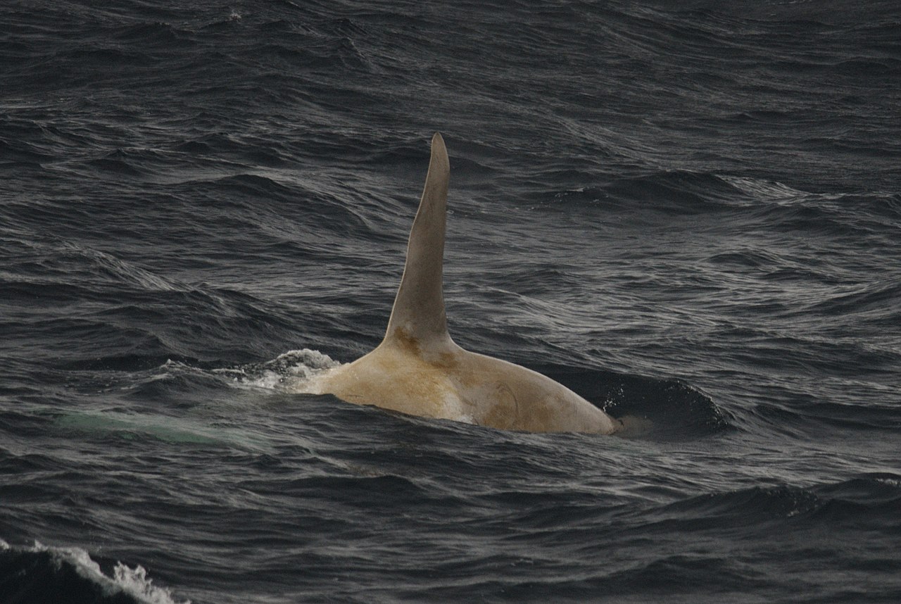
Diseases comparable to the Chédiak-Higashi-Syndrome (CHS) and the Hermansky-Pudlak-Syndrome (HPS) exist in the mouse, the Aleut mink, the Persian cat, the cattle, the pig, the rat, the fox and even the killer whale.
Cyclic hematopoiesis of the dog also resembles these syndromes. Affected animals are silver-grey, the number of neutrophil granulocytes is periodically reduced, as are red blood cells (erythrocytopenia) and platelets (thrombocytopenia). This leads to a blood clotting disorder and increased susceptibility to infections. Mostly the animals die shortly after birth.


Questions and answers
Q: What is albinism?
A: Albinism is a condition characterized by a lack of pigment melanin in hair, eyes, and skin, which can occur in people, animals, and plants.
Q: What causes albinism?
A: Albinism is caused by the absence or reduction of melanin, which is responsible for skin, hair and eye colour.
Q: What are some physical characteristics of people with albinism?
A: People with albinism often have very pale skin, white or light blonde hair, and blue or pinkish eyes.
Q: What are some problems that people with albinism can experience?
A: People with albinism can experience vision problems, such as nystagmus, strabismus, and refractive errors, as well as easily getting sunburnt due to a lack of melanin in the skin.
Q: How rare is albinism in the United States?
A: Albinism is rare in the United States, with only one out of every 20,000 people in the United States having the condition.
Q: How many people in the United States have albinism?
A: There are about 15,945 people in the United States who have albinism.
Q: What are some disadvantages that albino animals may experience?
A: Albino animals lack the camouflage that non-albino members of their species have and are more easily seen by predators. Also, where colour is a factor in mate selection, they may be at a disadvantage too.
Related articles
Author
AlegsaOnline.com Albinism: causes, characteristics, and impacts in humans, animals, and plants Leandro Alegsa
URL: https://en.alegsaonline.com/art/2154
Sources
- albinism.org : "What is Albinism"
- galenet.galegroup.com : "Albinism"