Bronchoscopy: examination and treatment of the airways
Bronchoscopy is an endoscopic procedure for visualizing and treating the trachea, bronchi and bronchioles. Methods include rigid, flexible fiberoptic and CT virtual bronchoscopy for diagnosis and interventions.
Overview
Bronchoscopy is a medical procedure that allows clinicians to view and access the internal surfaces of the large airways. Using specialized scopes, practitioners examine the trachea, main airways, and branching passages such as the bronchi and smaller bronchioles. It serves both diagnostic and therapeutic roles: identifying lesions, sampling tissue or fluid, and treating obstructing lesions or foreign bodies.
Image gallery
7 Images


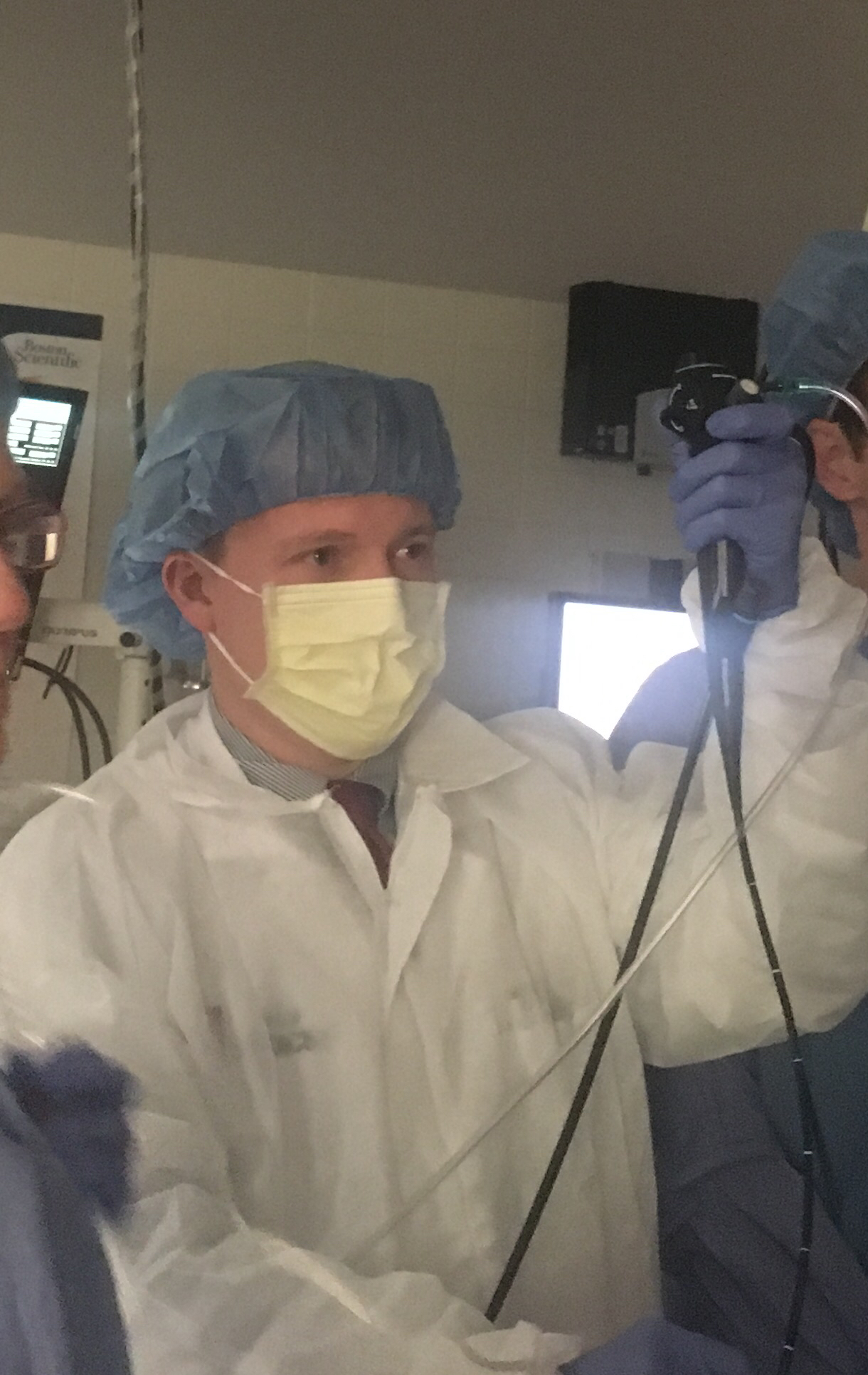

Types and equipment
There are three principal approaches in routine use. Rigid bronchoscopy employs a straight metal tube and is typically performed under general anesthesia; it remains important for removing large foreign bodies and placing airway stents. Flexible fiberoptic bronchoscopy uses a bendable scope allowing access to more distal airways and is commonly used with local anesthesia and sedation. CT virtual bronchoscopy is a noninvasive imaging technique that reconstructs airway views from computed tomography data and can guide planning though it does not permit interventions.
Procedure and common interventions
Before the procedure, patients are assessed for airway risk and bleeding tendency. During bronchoscopy a clinician may perform one or more of the following:
- direct inspection of airway anatomy and mucosa;
- endobronchial biopsy or transbronchial biopsy to sample tissue;
- removal of secretions, mucus plugs or aspirated foreign objects;
- placement of stents or dilatation for narrowed airways, and control of bleeding.
Indications and examples
Bronchoscopy is used when imaging or symptoms suggest airway disease. Typical reasons include investigating persistent cough, unexplained haemoptysis (coughing blood), abnormal chest imaging, suspected infection that requires microbiology, staging or diagnosing lung cancer, and assessing airway injury or obstruction. In intensive care settings it can help manage secretions and support ventilation strategies.
Safety, risks and limitations
The procedure is generally safe when performed by trained teams, but risks exist. Possible complications include bleeding after biopsy, pneumothorax with transbronchial sampling, infection, oxygen desaturation, and airway spasm. Contraindications or cautions include severe uncontrolled bleeding disorders, unstable cardiovascular status, or inadequate oxygenation that cannot be corrected. CT virtual bronchoscopy avoids many procedural risks but cannot replace sampling or therapeutic maneuvers.
History and development
Bronchoscopy evolved from rigid instruments developed in earlier centuries to flexible fiberoptic scopes in the twentieth century, which greatly expanded reach and comfort. Advances in imaging and computed tomography led to virtual bronchoscopy, improving preprocedural planning and noninvasive assessment. Ongoing improvements in optics, sampling tools, and interventional devices continue to broaden bronchoscopic capabilities.
Notable distinctions
When choosing a modality, clinicians weigh the need for tissue sampling or intervention against patient stability and available expertise. Rigid techniques excel when large-bore access or heavy bleeding control is needed; flexible scopes are preferred for outpatient diagnostics and finer sampling; CT virtual bronchoscopy is valuable for mapping anatomy and complementing direct visualization.
For further clinical guidelines and technical details, consult specialty resources and institutional protocols. Anatomy references and clinical procedure guides can help clinicians and learners contextualize bronchoscopic findings.
Questions and answers
Q: What is bronchoscopy?
A: Bronchoscopy is a medical procedure that is used to examine the inside of a person's airways.
Q: What is the purpose of bronchoscopy?
A: The purpose of bronchoscopy is to see the inside of the trachea, bronchi, and bronchioles within the lungs.
Q: What are the different types of bronchoscopy?
A: The different types of bronchoscopy are rigid, flexible fiberoptic bronchoscopy, and CT virtual bronchoscopy.
Q: What is rigid bronchoscopy?
A: Rigid bronchoscopy is a type of bronchoscopy that uses a straight, inflexible tube to look inside the airways.
Q: What is flexible fiberoptic bronchoscopy?
A: Flexible fiberoptic bronchoscopy is a type of bronchoscopy that uses a thin, flexible tube with a camera and light on the end to examine the airways.
Q: What is CT virtual bronchoscopy?
A: CT virtual bronchoscopy is a type of bronchoscopy that uses computerized tomography (CT) scans to create a 3D image of the airways.
Q: Why is bronchoscopy conducted?
A: Bronchoscopy is conducted to diagnose respiratory problems, such as lung cancer, infections, inflammation, and blockages in the airways.
Related articles
Author
AlegsaOnline.com Bronchoscopy: examination and treatment of the airways Leandro Alegsa
URL: https://en.alegsaonline.com/art/14672