
X-ray crystallography: determining molecular arrangements by diffraction
A concise overview of X-ray crystallography: how it reveals atomic arrangements, the physical principles, historical milestones, methods, applications in chemistry and biology, and key limitations.
Overview
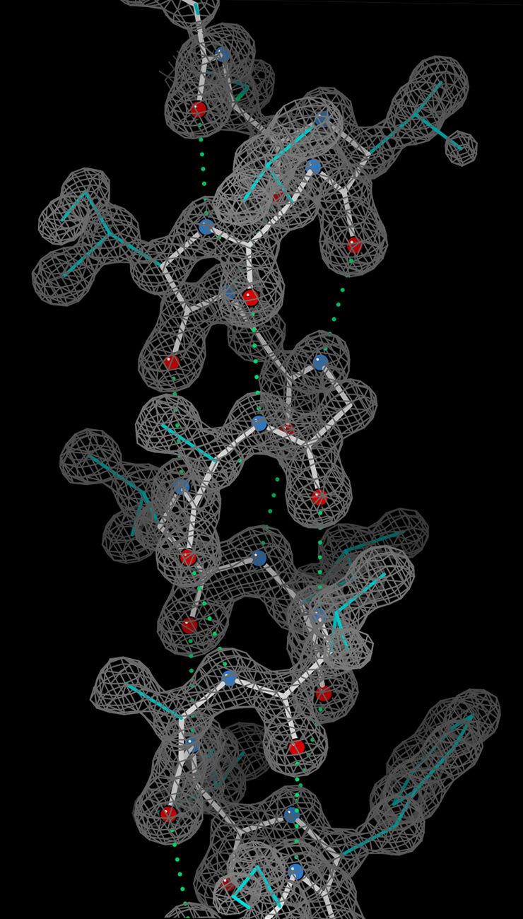
X-ray crystallography is a laboratory technique used to determine the precise three-dimensional arrangement of atoms in crystalline matter. By analyzing patterns produced when a beam of X-rays strikes a crystal, researchers generate maps of electron density and infer the positions of atoms to build models of the three-dimensional structure and overall molecular structure. The method applies to both small chemical compounds and large biological macromolecules, and it underpins structural chemistry, materials science and structural biology.
Image gallery
10 Images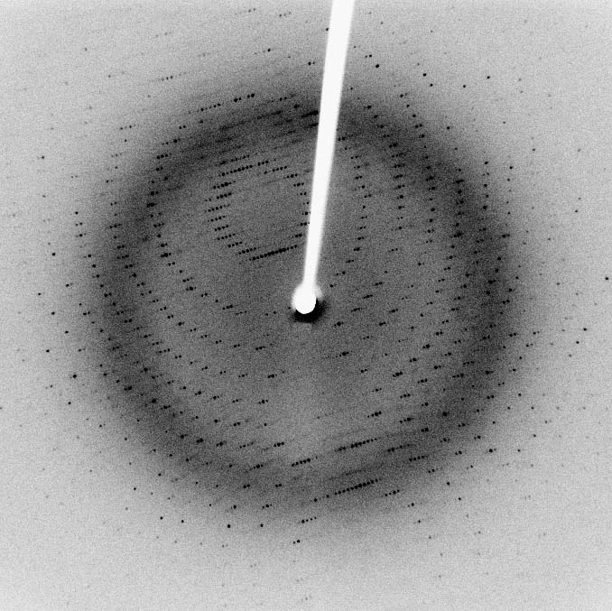





Physical principles and key steps
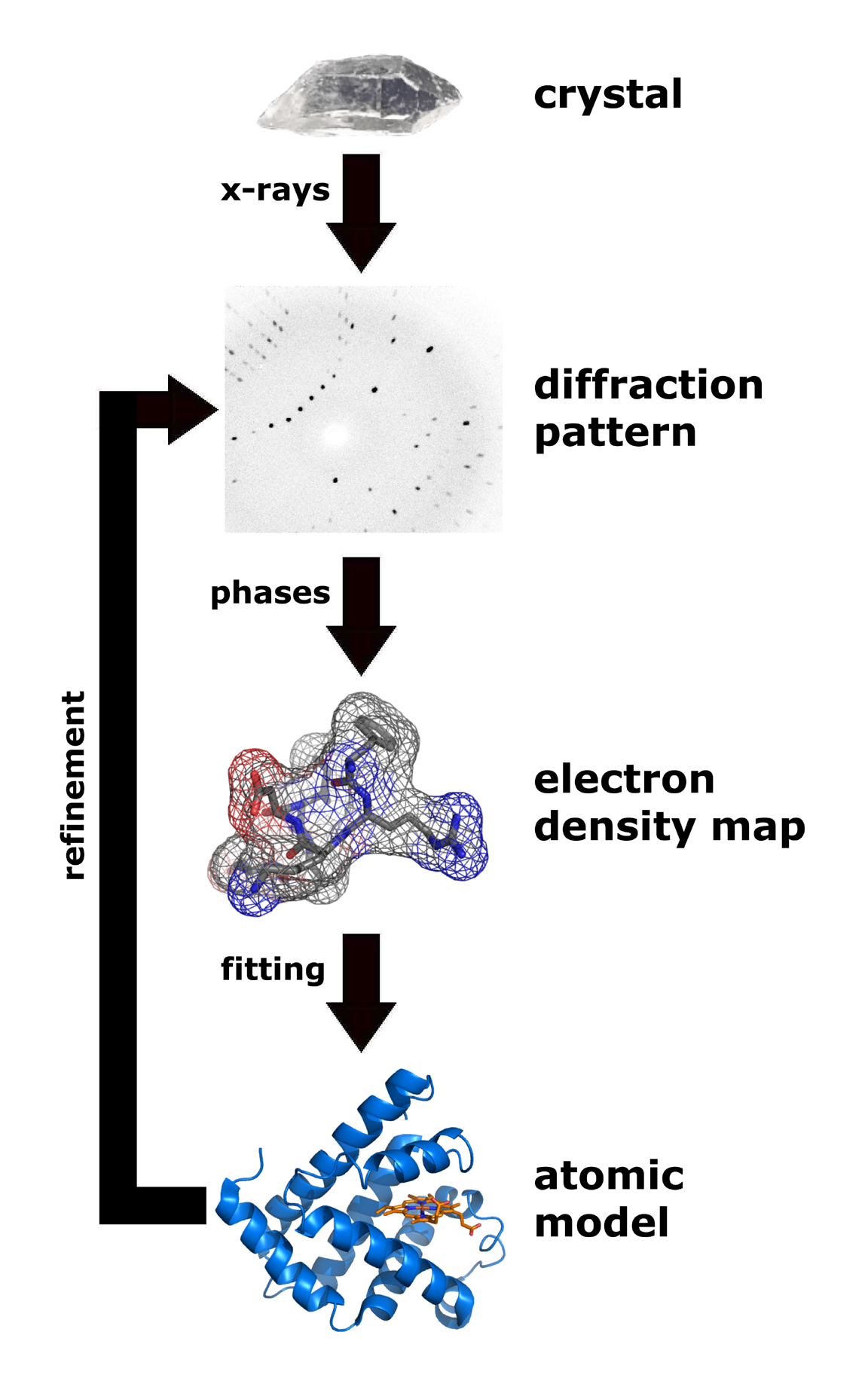
X-rays interact primarily with the electron cloud surrounding each atom, so scattering intensities reflect electron density. A crystal organizes molecules or atoms into a repeating lattice so scattered waves interfere and form a diffraction pattern. Typical workflow steps are:
- grow a suitable crystal of the target molecule or material,
- collect diffraction images using laboratory sources or synchrotron beamlines,
- process intensities and solve the phase problem to produce an electron-density map,
- build and refine an atomic model that fits the density.
History and milestones
The foundations of the method were formalized in the early 20th century by Sir William and Sir Lawrence Bragg, who related diffraction patterns to lattice planes and established crystallography as a quantitative science. Their work earned the 1915 Nobel Prize in Physics. X-ray studies later enabled the discovery of the DNA double helix and many protein structures, with contributions from researchers such as Watson, Crick, Wilkins and Rosalind Franklin, who provided critical experimental data.
Methods and variants
The classical approach is single-crystal X-ray diffraction, where a single well-ordered crystal yields high-resolution information. Powder diffraction (XRD) studies polycrystalline or powdered samples and is widely used for phase identification and materials characterization. Modern facilities use cryo-cooling to reduce radiation damage, and powerful synchrotron sources or X-ray free-electron lasers provide intense, tunable beams that extend capabilities to tiny crystals and time-resolved experiments.
Applications and examples
X-ray crystallography has immense practical impact. In chemistry it confirms bond lengths, stereochemistry and conformations of organic compounds, while in materials science it reveals atomic arrangements of inorganic materials and catalysts. In biology it determines protein, nucleic acid and complex assemblies structures, guiding drug development and illuminating mechanisms of enzymes, receptors and transporters. Structural databases created from crystallographic models are essential resources for research and industry.
Limitations and notable facts
The technique requires a crystalline sample and may not capture flexible or heterogeneous regions well; some molecules resist crystallization. Radiation damage, model bias and interpretation of ambiguous density are practical concerns. Despite these limits, X-ray crystallography remains a gold standard for atomic-resolution structure determination and continues to evolve with advances in sample preparation, instrumentation and computation.
Questions and answers
Q: What is X-ray crystallography?
A: X-ray crystallography is a technique used to see the three-dimensional structure of a molecule, which creates a picture on a screen by bending X-rays from the electron cloud of an atom.
Q: Can X-ray crystallography be used for both organic and inorganic molecules?
A: Yes, X-ray crystallography can be used to study both organic and inorganic molecules.
Q: Who are the inventors of X-ray crystallography?
A: Sir William Bragg and his son Sir Lawrence Bragg jointly invented X-ray crystallography and won the Nobel Prize in Physics in 1915 for their discovery.
Q: What is the oldest method of X-ray crystallography?
A: The oldest method of X-ray crystallography is X-ray diffraction (XRD), where X-rays are fired at a single crystal to produce a pattern that can be used to determine the arrangement of atoms inside the crystal.
Q: Was the sample destroyed during the X-ray crystallography process?
A: No, the sample is not destroyed during the X-ray crystallography process.
Q: Who was the Director of the Cavendish Laboratory when the discovery of the structure of DNA was made?
A: Sir Lawrence Bragg was the Director of the Cavendish Laboratory, Cambridge University, when the discovery of the structure of DNA was made by James D. Watson, Francis Crick, Maurice Wilkins, and Rosalind Franklin in February 1953.
Q: Who is the youngest Nobel Laureate in Physics?
A: Sir Lawrence Bragg is the youngest Nobel Laureate in Physics, having won the award in 1915 for his joint discovery of X-ray crystallography with his father Sir William Bragg.
Related articles
Author
AlegsaOnline.com X-ray crystallography: determining molecular arrangements by diffraction Leandro Alegsa
URL: https://en.alegsaonline.com/art/109413
Sources
- panalytical.com : "Introduction to X-ray Diffraction (XRD)"