
Wernicke's area — cortical region for language comprehension
Wernicke's area is a region in the dominant posterior temporal cortex involved in understanding spoken and written language; damage leads to fluent but often meaningless speech and impaired comprehension.
Wernicke's area is a region of the brain traditionally associated with comprehension of language and with the mapping of sounds and visual symbols onto meaning. Many overviews and resources refer to its role in receptive language processing; see a general summary at Wernicke's area. The region is commonly discussed alongside Broca's area, which is linked more strongly to production and motor aspects of speech.
Image gallery
1 Image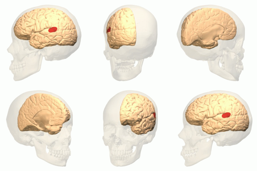
Anatomy and lateralization
Classical anatomical descriptions place Wernicke's area in the posterior portion of the superior temporal gyrus, often corresponding roughly to Brodmann area 22, in the dominant hemisphere. It lies adjacent to primary auditory cortex and the planum temporale and forms part of the larger temporal lobe language zone; for broader neuroanatomical context, see resources on the cerebral cortex and the temporal lobe. Language dominance is lateralized in most people: the left hemisphere is dominant in about 95% of right-handed individuals and in a majority of left-handed people, though individual variability is common.
Functions and processing
Contemporary models emphasize that Wernicke's area contributes to several aspects of language processing rather than serving a single narrowly defined function. It is important for mapping incoming auditory speech and visual word forms to lexical and conceptual representations, for supporting lexical access and word meaning, and for integrating semantic and syntactic information so that sentences are understood. The area participates in networks for both spoken and written language and interacts with frontal, parietal and subcortical regions as part of distributed processing pathways.
Clinical features and aphasia
Lesions affecting this cortical region produce a syndrome historically called Wernicke's aphasia or receptive aphasia. Patients typically produce fluent, well formed sentences that may be semantically empty, with frequent word-finding errors, paraphasias (substituted or distorted words), and newly formed words (neologisms). Comprehension of spoken and written language is markedly impaired, and repetition is often disrupted. In severe cases speech may appear as a jumble of words and phrases with little coherent meaning, a phenomenon sometimes described as "word salad".
- Common signs: fluent but nonsensical speech, impaired comprehension, anomia (difficulty naming objects).
- Associated impairments: poor repetition, reading and writing difficulties that reflect disrupted comprehension rather than purely motor deficits.
Assessment and differential diagnosis
Clinical assessment of suspected receptive aphasia includes testing spontaneous speech, naming, comprehension of single words and sentences, repetition, and reading. Distinguishing damage to Wernicke's cortex from disconnection syndromes (for example conduction aphasia, where the pathway between comprehension and production areas is disrupted) or from broader semantic memory disorders is important for prognosis and management. Neuroimaging and careful bedside testing help localize the deficit and characterize its nature.
Connections and network perspective
Wernicke's area does not act in isolation. It is interconnected with frontal speech areas, including Broca's region, via fiber bundles such as the arcuate fasciculus and other dorsal and ventral language pathways revealed by modern tractography. Contemporary neuroscience favors network models in which comprehension arises from coordinated activity across temporal, parietal and frontal cortices, supported by subcortical structures.
History and clinical relevance
The area is named after Carl Wernicke, a 19th‑century German neurologist who related a characteristic pattern of comprehension impairment to lesions in the posterior temporal lobe after careful clinical observation and postmortem study; his findings complemented Paul Broca's earlier work on expressive language and helped establish clinicopathological correlation in neurology. Early conclusions were based on autopsies of patients with language deficits. Today, Wernicke's area remains a central concept in neurology, neuropsychology and speech‑language pathology for localizing and treating language disorders.
Imaging, rehabilitation and research
Functional imaging (fMRI, PET) and modern lesion studies show that activity related to comprehension extends beyond the classical Wernicke site, involving adjacent temporal and parietal regions and bilateral contributions in some tasks. Rehabilitation after injury includes speech and language therapy focused on improving comprehension, compensatory strategies, and retraining lexical access; recovery depends on lesion size, location, age, and therapy intensity. Ongoing research explores how distributed networks, neuroplasticity and interhemispheric reorganization contribute to recovery and to individual differences in language organization.
For accessible introductions to speech and language systems, see entries on speech and regional overviews at Wernicke's area. Historical and clinical descriptions that originally linked language deficits to localized cortical lesions continue to inform modern, network-based perspectives on how the brain supports meaning, hearing and communication.
Questions and answers
Q: What is Wernicke's area?
A: Wernicke's area is an area of the brain located in Brodmann area 22, in the dominant cerebral hemisphere. It is one of two parts of the cerebral cortex linked to speech (the other being Broca's area). It is involved in the production and comprehension of written and spoken language.
Q: How was Wernicke's area discovered?
A: Wernicke's area was discovered by autopsies of patients who had speech difficulties. Carl Wernicke, a German neurologist, had a patient who could speak quite well but was unable to understand the speech of others. After this patient passed away, it was found that there was damage to an area at the upper portion of the temporal lobe just behind the auditory cortex which led to its discovery.
Q: What type of aphasia does this part of the brain cause?
A: Damage to Wernicke's area can lead to what is known as "Wernicke's aphasia" or "receptive aphasia". This type of aphasia causes people difficulty understanding language and responding with sentences that are grammatical but make little sense - often referred to as "word salad".
Q: Does this part of the brain only affect speech comprehension?
A: No, damage to Wernicke's Area can also lead to difficulty naming things - often responding with words that sound similar or related names instead.
Q: Where are Broca’s and Wernicke’s areas located relative to each other?
A: Despite being located in different lobes, Broca’s and Wernicke’s areas are quite close together. They are connected by nerves called arcuate fascilicus as well as another recently discovered tract.
Q: In what percentage of right-handed individuals is their dominant hemisphere on their left side?
A: In about 95% percent right-handed individuals, their dominant hemisphere will be on their left side. For left-handed individuals it would be around 60%.
Related articles
Author
AlegsaOnline.com Wernicke's area — cortical region for language comprehension Leandro Alegsa
URL: https://en.alegsaonline.com/art/107303
Sources
- webspace.ship.edu : "Speech and the brain"
- onlinelibrary.wiley.com : onlinelibrary.wiley.com/doi/10.1111/j.1749-6632.1976.tb25546.x/abstract?systemMessage=Wil…
- asha.org : "Aphasia: signs & symptoms"
- onlinelibrary.wiley.com : onlinelibrary.wiley.com/doi/10.1002/ana.20319/abstract?systemMessage=Wiley+Online+Library…