
Vomer bone
Thin, unpaired facial bone forming the lower and posterior part of the nasal septum; contributes to the separation of the two nasal cavities and has clinical importance in nasal structure and surgery.
The vomer is a thin, unpaired bone positioned along the midline of the nasal cavity. It forms the lower and rear portion of the nasal septum, the wall that divides the two nasal passages. The term vomer comes from a Latin word meaning "plowshare," a reference to its flattened, blade-like shape. Anatomically it is a small but central element in the structure of the nose and midface; see vomer for contextual diagrams and references.
Image gallery
10 Images
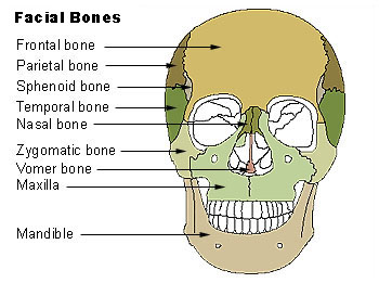


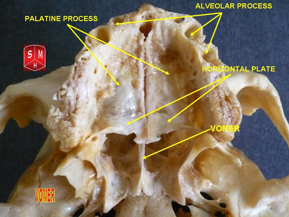

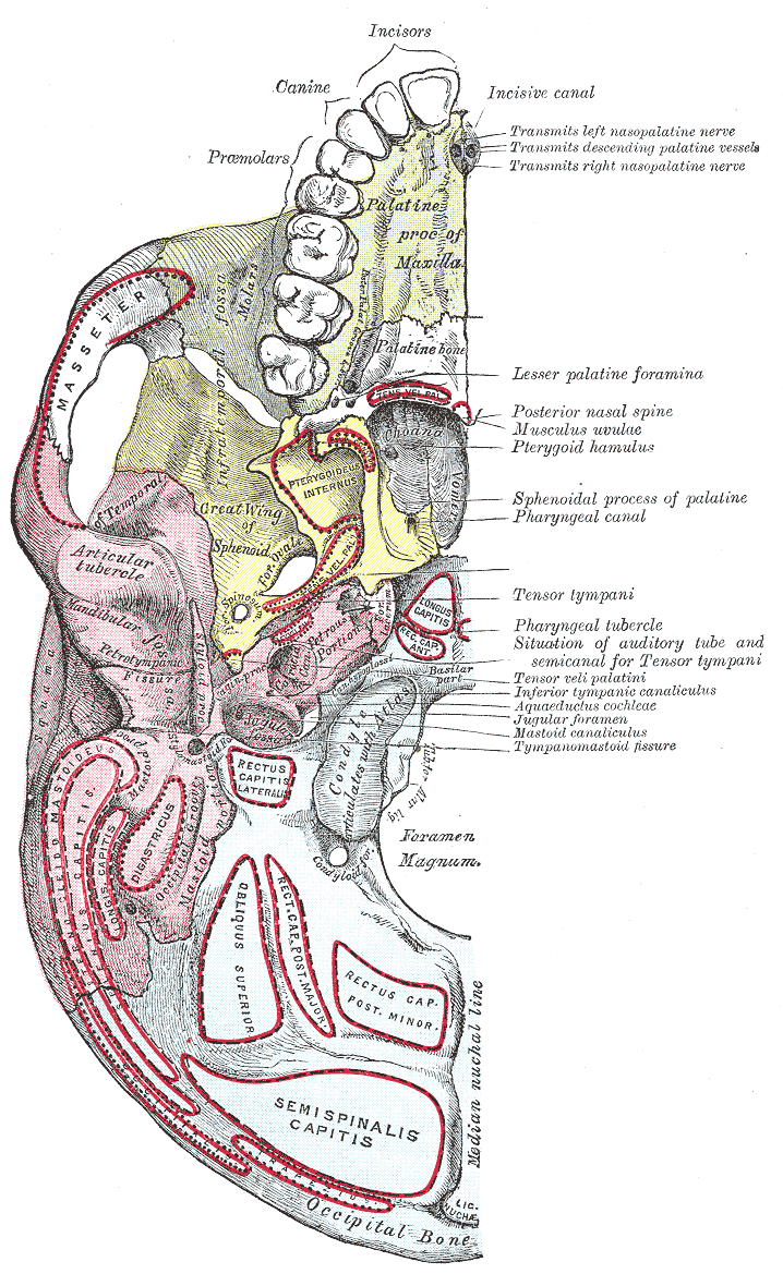


Structure and characteristics
The vomer is generally trapezoidal in outline, thin and plate-like. It occupies the posteroinferior part of the septal framework, lying below the perpendicular plate of the ethmoid and behind the septal cartilage. It is unpaired and located in the median plane, contributing a rigid bony surface to support the soft tissues of the septum.
Articulations and relations
- Superiorly it contacts parts of the sphenoid and the perpendicular plate of the ethmoid.
- Inferiorly it rests against the maxilla and palatine bones and helps form the nasal crest.
- Anteroinferiorly it is continuous with the septal cartilage and mucosal lining.
Because the vomer sits amid several facial bones, variations in its shape or position can affect overall nasal form; it is described alongside other facial bones in regional studies of the face.
Development and etymology
The vomer begins as ossified tissue during fetal development and typically arises from distinct centers that unite as growth proceeds. Its Latin-derived name—meaning plowshare—reflects the long-recognized resemblance to an agricultural blade. The bone has been described in classical anatomical texts and remains a consistent landmark in cranial anatomy.
Function and clinical significance
Functionally, the vomer helps maintain separation between the two nasal cavities, stabilizes the septal cartilage, and contributes to normal airflow and mucosal support. Clinically, deviations, fractures or congenital variations of the vomer can contribute to a deviated nasal septum, nasal obstruction, or complications in septal surgery. Surgeons performing septoplasty or reconstructive procedures take vomer anatomy into account to restore airway function.
Notable facts and distinctions
Unlike paired facial bones, the vomer is singular and centrally positioned. It forms the bony posterior-inferior part of the septum together with the perpendicular plate of the ethmoid and the septal cartilage. Its relationships to the surrounding skull are often illustrated in cranial overviews and osteological collections; further anatomical context is available in skull references such as cranial anatomy.
Questions and answers
Q: What is the vomer?
A: The vomer is a thin, unpaired bone of the face and skull (cranium) located in the center of the nasal cavity.
Q: Where is the vomer located?
A: The vomer is located in the center of the nasal cavity.
Q: What shape is the vomer?
A: The vomer is trapezoidal in shape.
Q: What does the vomer do?
A: The vomer serves as part of the nasal septum, which is the middle wall of the nasal respiratory cavity.
Q: Is it a paired bone?
A: No, it is an unpaired bone.
Q: Is it a large or small bone?
A: It is a small bone.
Q: What other bones make up part of the nasal septum?
A: Other bones that make up part of the nasal septum include ethmoid and maxillary bones.
Related articles
Author
AlegsaOnline.com Vomer bone Leandro Alegsa
URL: https://en.alegsaonline.com/art/105909