Trachea (windpipe): structure, function and clinical significance
The trachea, or windpipe, is the sturdy, cartilaginous airway that conducts air from the larynx to the lungs. This article explains its anatomy, histology, development and common clinical issues.
Overview
The trachea, commonly called the windpipe, is a tubular airway that conveys air between the throat and the lungs. In vertebrates it forms a central part of the respiratory system, providing a low-resistance path for inhaled and exhaled air. In humans and other mammals, the trachea begins at the lower edge of the larynx and continues down to the point where it divides into the left and right primary bronchi that enter the lungs.
Image gallery
10 Images


Anatomy and microscopic structure
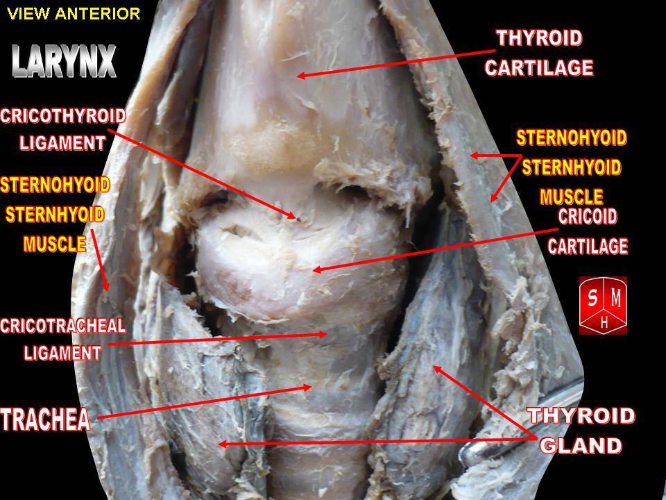
Macroscopically the trachea is a flexible tube supported by a series of C-shaped cartilage rings that keep it open while permitting movement and slight compression. The back wall of the trachea is completed by a band of muscle and connective tissue, which allows the esophagus to expand during swallowing. Typical adult tracheal length is modest (often around 10–13 cm), subject to individual variation.
On the inside, the tracheal lining is made of ciliated pseudostratified columnar epithelium with goblet cells. Cilia and mucus trap and move inhaled particles upward toward the throat, helping to protect the lower airways. The point where the trachea splits is called the carina, an important landmark in anatomy and in procedures such as bronchoscopy.
Development and comparative notes
The trachea develops embryologically from the foregut, separating as a distinct tube early in gestation. Its detailed form varies across the animal kingdom: for example, some vertebrates have more rigid or differently supported airways, but the basic role of conducting air is conserved. See general vertebrate comparisons here.
Functions and importance
- Air conduction: provides an open channel for breathing between the upper airway and the lungs.
- Air conditioning and defense: cilia and mucus humidify and cleanse inhaled air.
- Protection of lower airway: cartilage and reflexes (like coughing) help keep the trachea patent and clear.
Clinical relevance
The trachea can be affected by infections, inflammation and structural problems. Tracheitis refers to inflammation of the tracheal mucosa, which may accompany upper respiratory infections. Other conditions include tracheal stenosis (narrowing), tracheomalacia (loss of structural support) and foreign-body obstruction. Medical and surgical interventions range from endoscopic evaluation to procedures such as tracheostomy to secure an airway when needed.
Practical and procedural notes
Healthcare providers often examine the trachea with imaging and endoscopy when patients present with unexplained cough, breathing difficulty or suspected obstruction. The relationship of the trachea to adjacent structures—such as the thyroid gland, esophagus and major blood vessels—is important during neck surgery and emergency airway management. For basic patient information about the nose and mouth as entry points to the trachea, consult resources on the nose and mouth. For a general introduction to airway anatomy and procedures see this overview and clinical guidance on bronchoscopy and airway care here or here.
Questions and answers
Q: What is the trachea?
A: The trachea, or windpipe, is the bony tube that connects the nose and mouth to the lungs.
Q: Where does the trachea begin and end in mammals?
A: In mammals, the trachea begins at the lower part of the larynx and continues to the lungs.
Q: What happens to the trachea once it reaches the lungs?
A: The trachea branches into the right and left bronchi once it reaches the lungs.
Q: What is tracheitis?
A: Tracheitis is the inflammation of the linings of the trachea.
Q: Can inflammation of the trachea lead to other conditions?
A: Yes, inflammation of the trachea can lead to other conditions, such as tracheitis.
Q: How important is the trachea to the vertebrate respiratory system?
A: The trachea is an important part of the vertebrate respiratory system.
Q: Is the trachea the same thing as the windpipe?
A: Yes, the trachea is the same thing as the windpipe.
Related articles
Author
AlegsaOnline.com Trachea (windpipe): structure, function and clinical significance Leandro Alegsa
URL: https://en.alegsaonline.com/art/100998
Sources
- doi.org : 10.1126/science.1078008
- pubmed.ncbi.nlm.nih.gov : 12543973